- PDB-1sor: Aquaporin-0 membrane junctions reveal the structure of a closed w... -
+
Open data
ID or keywords:
Loading...
-
Basic information
Entry
Database: PDB / ID: 1sor
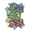
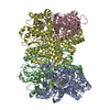
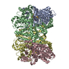
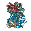
Title
Aquaporin-0 membrane junctions reveal the structure of a closed water pore
Components
Aquaporin-0
Keywords
MEMBRANE PROTEIN / membrane junction / water channel
Function / homology
Function and homology information
gap junction-mediated intercellular transport / water transport / water channel activity / structural constituent of eye lens / gap junction / lens development in camera-type eye / positive regulation of cell adhesion / visual perception / protein homotetramerization / calmodulin binding ...gap junction-mediated intercellular transport / water transport / water channel activity / structural constituent of eye lens / gap junction / lens development in camera-type eye / positive regulation of cell adhesion / visual perception / protein homotetramerization / calmodulin binding / apical plasma membrane / endoplasmic reticulum / plasma membrane Similarity search - Function
Glycerol uptake facilitator protein / Glycerol uptake facilitator protein. / Aquaporin transporter / Major intrinsic protein, conserved site / MIP family signature. / Major intrinsic protein / Major intrinsic protein / Aquaporin-like / Up-down Bundle / Mainly Alpha Similarity search - Domain/homology
Journal: Nature / Year: 2004 Title: Aquaporin-0 membrane junctions reveal the structure of a closed water pore. Authors: Tamir Gonen / Piotr Sliz / Joerg Kistler / Yifan Cheng / Thomas Walz / Abstract: The lens-specific water pore aquaporin-0 (AQP0) is the only aquaporin known to form membrane junctions in vivo. We show here that AQP0 from the lens core, containing some carboxy-terminally cleaved ...The lens-specific water pore aquaporin-0 (AQP0) is the only aquaporin known to form membrane junctions in vivo. We show here that AQP0 from the lens core, containing some carboxy-terminally cleaved AQP0, forms double-layered crystals that recapitulate in vivo junctions. We present the structure of the AQP0 membrane junction as determined by electron crystallography. The junction is formed by three localized interactions between AQP0 molecules in adjoining membranes, mainly mediated by proline residues conserved in AQP0s from different species but not present in most other aquaporins. Whereas all previously determined aquaporin structures show the pore in an open conformation, the water pore is closed in AQP0 junctions. The water pathway in AQP0 also contains an additional pore constriction, not seen in other known aquaporin structures, which may be responsible for pore gating.
History
Deposition
Mar 15, 2004
Deposition site: RCSB / Processing site: RCSB
Revision 1.0
May 11, 2004
Provider: repository / Type: Initial release
Revision 1.1
Apr 29, 2008
Group: Version format compliance
Revision 1.2
Jul 13, 2011
Group: Derived calculations / Version format compliance
Revision 1.3
Oct 11, 2017
Group: Data collection / Data processing / Refinement description Category: em_3d_reconstruction / em_image_scans / software
EXPERIMENT TYPE : SINGLE-CRYSTAL ELECTRON DIFFRACTION DATE OF DATA COLLECTION : 28-JAN-2003 ...EXPERIMENT TYPE : SINGLE-CRYSTAL ELECTRON DIFFRACTION DATE OF DATA COLLECTION : 28-JAN-2003 TEMPERATURE (KELVIN) : 100.0 PH : 6.00 NUMBER OF CRYSTALS USED : 131 RADIATION SOURCE : ELECTRON MICROSCOPE X-RAY GENERATOR MODEL : TECNAI T20 OPTICS : CRYSTALS TILTED TO 0, 20, 45, 60 AND 70 DEGREES DETECTOR TYPE : CCD DETECTOR MANUFACTURER : GATAN 2K X 2K INTENSITY-INTEGRATION SOFTWARE : DIGITAL MICROGRAPH 3.7.4 DATA SCALING SOFTWARE : MRC NUMBER OF UNIQUE REFLECTIONS : 6635 RESOLUTION RANGE HIGH (A) : 3.000 RESOLUTION RANGE LOW (A) : 30.000 VERALL. COMPLETENESS FOR RANGE (%) : 88.0 DATA REDUNDANCY : 6.700 IN THE HIGHEST RESOLUTION SHELL. HIGHEST RESOLUTION SHELL, RANGE HIGH (A) : 3.00 HIGHEST RESOLUTION SHELL, RANGE LOW (A) : 3.50 COMPLETENESS FOR SHELL (%) : 82.0 DATA REDUNDANCY IN SHELL : 4.50 R MERGE FOR SHELL (I) : 0.54000 METHOD USED TO DETERMINE THE STRUCTURE: MOLECULAR REPLACEMENT SOFTWARE USED: MOLREP 7.4.03 STARTING MODEL: PDB ENTRY 1J4N
Remark 999
SEQUENCE The sequence of this protein has been deposited to gene bank. The accession number is AY573927.
-
Structure visualization
Movie
Biological unit as author_and_software_defined_assembly
In the structure databanks used in Yorodumi, some data are registered as the other names, "COVID-19 virus" and "2019-nCoV". Here are the details of the virus and the list of structure data.
Jan 31, 2019. EMDB accession codes are about to change! (news from PDBe EMDB page)
EMDB accession codes are about to change! (news from PDBe EMDB page)
The allocation of 4 digits for EMDB accession codes will soon come to an end. Whilst these codes will remain in use, new EMDB accession codes will include an additional digit and will expand incrementally as the available range of codes is exhausted. The current 4-digit format prefixed with “EMD-” (i.e. EMD-XXXX) will advance to a 5-digit format (i.e. EMD-XXXXX), and so on. It is currently estimated that the 4-digit codes will be depleted around Spring 2019, at which point the 5-digit format will come into force.
The EM Navigator/Yorodumi systems omit the EMD- prefix.
Related info.:Q: What is EMD? / ID/Accession-code notation in Yorodumi/EM Navigator
Yorodumi is a browser for structure data from EMDB, PDB, SASBDB, etc.
This page is also the successor to EM Navigator detail page, and also detail information page/front-end page for Omokage search.
The word "yorodu" (or yorozu) is an old Japanese word meaning "ten thousand". "mi" (miru) is to see.
Related info.:EMDB / PDB / SASBDB / Comparison of 3 databanks / Yorodumi Search / Aug 31, 2016. New EM Navigator & Yorodumi / Yorodumi Papers / Jmol/JSmol / Function and homology information / Changes in new EM Navigator and Yorodumi
 Movie
Movie Controller

 Yorodumi
 Open data
 Basic information
 Components
 Keywords
 Function and homology information
 MOLECULAR REPLACEMENT / cryo EM / Resolution: 3 Å
  Authors
 Citation
 Structure visualization
 Downloads & links
 Other downloads
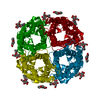


 PDBj
PDBj
 Assembly
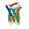

 Sample preparation
 Processing