Movie
Movie Controller

[English] 日本語
 Yorodumi
- EMDB-2355: Negative staining three dimensional reconstruction of bacteriopha... -
+ Open data

- Basic information
| Entry | Database: EMDB / ID: EMD-2355 | |||||||||
|---|---|---|---|---|---|---|---|---|---|---|
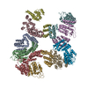
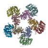
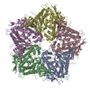
| Title | Negative staining three dimensional reconstruction of bacteriophage T7 large terminase | |||||||||
 Map data Map data | Threedimensional reconstruction of bacteriophage T7 large terminase | |||||||||
 Sample Sample |
| |||||||||
 Keywords Keywords | ATPase / bacteriophage / DNA translocation / Packaging / single-particle reconstruction / terminase. | |||||||||
| Function / homology |  Function and homology information Function and homology information: / viral terminase, large subunit / viral DNA genome packaging / nuclease activity / Hydrolases; Acting on ester bonds; Endodeoxyribonucleases producing 5'-phosphomonoesters / chromosome organization / Hydrolases; Acting on acid anhydrides; Acting on acid anhydrides to facilitate cellular and subcellular movement / endonuclease activity / nucleotide binding / ATP hydrolysis activity ...: / viral terminase, large subunit / viral DNA genome packaging / nuclease activity / Hydrolases; Acting on ester bonds; Endodeoxyribonucleases producing 5'-phosphomonoesters / chromosome organization / Hydrolases; Acting on acid anhydrides; Acting on acid anhydrides to facilitate cellular and subcellular movement / endonuclease activity / nucleotide binding / ATP hydrolysis activity / ATP binding / metal ion binding Similarity search - Function | |||||||||
| Biological species |   Enterobacteria phage T7 (virus) Enterobacteria phage T7 (virus) | |||||||||
| Method | single particle reconstruction / negative staining / Resolution: 16.0 Å | |||||||||
 Authors Authors | Dauden MI / Martin-Benito J / Sanchez-Ferrero JC / Pulido-Cid M / Valpuesta JM / Carrascosa JL | |||||||||
 Citation Citation | Journal: J Biol Chem / Year: 2013 Title: Large terminase conformational change induced by connector binding in bacteriophage T7. Authors: María I Daudén / Jaime Martín-Benito / Juan C Sánchez-Ferrero / Mar Pulido-Cid / José M Valpuesta / José L Carrascosa /  Abstract: During bacteriophage morphogenesis DNA is translocated into a preformed prohead by the complex formed by the portal protein, or connector, plus the terminase, which are located at an especial prohead ...During bacteriophage morphogenesis DNA is translocated into a preformed prohead by the complex formed by the portal protein, or connector, plus the terminase, which are located at an especial prohead vertex. The terminase is a powerful motor that converts ATP hydrolysis into mechanical movement of the DNA. Here, we have determined the structure of the T7 large terminase by electron microscopy. The five terminase subunits assemble in a toroid that encloses a channel wide enough to accommodate dsDNA. The structure of the complete connector-terminase complex is also reported, revealing the coupling between the terminase and the connector forming a continuous channel. The structure of the terminase assembled into the complex showed a different conformation when compared with the isolated terminase pentamer. To understand in molecular terms the terminase morphological change, we generated the terminase atomic model based on the crystallographic structure of its phage T4 counterpart. The docking of the threaded model in both terminase conformations showed that the transition between the two states can be achieved by rigid body subunit rotation in the pentameric assembly. The existence of two terminase conformations and its possible relation to the sequential DNA translocation may shed light into the molecular bases of the packaging mechanism of bacteriophage T7. | |||||||||
| History |
|
- Structure visualization
| Movie |
Movie viewer |
|---|---|
| Structure viewer | EM map: SurfViewMolmilJmol/JSmol |
| Supplemental images |

- Downloads & links
-EMDB archive
| Map data | emd_2355.map.gz | 1.8 MB | EMDB map data format | |
|---|---|---|---|---|
| Header (meta data) | emd-2355-v30.xmlemd-2355.xml | 10.7 KB 10.7 KB | Display Display | EMDB header |
| Images | 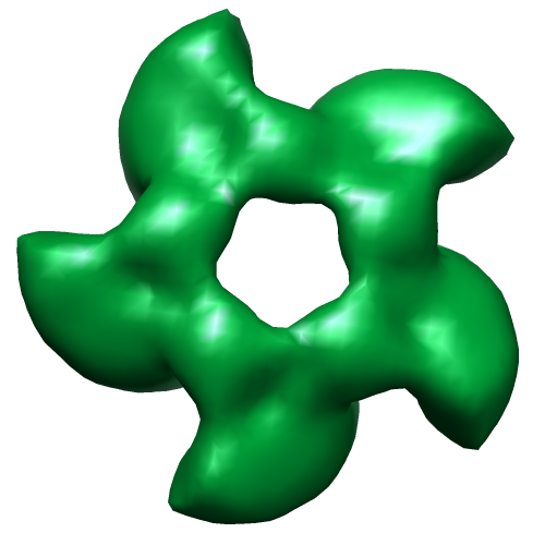 EMD-2355.png EMD-2355.png | 98.5 KB | ||
| Archive directory |  http://ftp.pdbj.org/pub/emdb/structures/EMD-2355ftp://ftp.pdbj.org/pub/emdb/structures/EMD-2355 http://ftp.pdbj.org/pub/emdb/structures/EMD-2355ftp://ftp.pdbj.org/pub/emdb/structures/EMD-2355 | HTTPS FTP |
-Validation report
| Summary document | emd_2355_validation.pdf.gz | 213 KB | Display | EMDB validaton report |
|---|---|---|---|---|
| Full document | emd_2355_full_validation.pdf.gz | 212.1 KB | Display | |
| Data in XML | emd_2355_validation.xml.gz | 4.7 KB | Display | |
| Arichive directory | https://ftp.pdbj.org/pub/emdb/validation_reports/EMD-2355ftp://ftp.pdbj.org/pub/emdb/validation_reports/EMD-2355 | HTTPS FTP |
-Related structure data
| Related structure data |  4bijMC  2356C  4bilC M: atomic model generated by this map C: citing same article ( |
|---|---|
| Similar structure data |

-Links
| EMDB pages | EMDB (EBI/PDBe) / EMDataResource |
|---|
-Map
| File | Download / File: emd_2355.map.gz / Format: CCP4 / Size: 1.9 MB / Type: IMAGE STORED AS FLOATING POINT NUMBER (4 BYTES) | ||||||||||||||||||||||||||||||||||||||||||||||||||||||||||||||||||||
|---|---|---|---|---|---|---|---|---|---|---|---|---|---|---|---|---|---|---|---|---|---|---|---|---|---|---|---|---|---|---|---|---|---|---|---|---|---|---|---|---|---|---|---|---|---|---|---|---|---|---|---|---|---|---|---|---|---|---|---|---|---|---|---|---|---|---|---|---|---|
| Annotation | Threedimensional reconstruction of bacteriophage T7 large terminase | ||||||||||||||||||||||||||||||||||||||||||||||||||||||||||||||||||||
| Voxel size | X=Y=Z: 4.4 Å | ||||||||||||||||||||||||||||||||||||||||||||||||||||||||||||||||||||
| Density |
| ||||||||||||||||||||||||||||||||||||||||||||||||||||||||||||||||||||
| Symmetry | Space group: 1 | ||||||||||||||||||||||||||||||||||||||||||||||||||||||||||||||||||||
| Details | EMDB XML:
CCP4 map header:
| ||||||||||||||||||||||||||||||||||||||||||||||||||||||||||||||||||||
-Supplemental data
- Sample components
-Entire : Full-length large terminase of bacteriophage T7
| Entire | Name: Full-length large terminase of bacteriophage T7 |
|---|---|
| Components |
|
-Supramolecule #1000: Full-length large terminase of bacteriophage T7
| Supramolecule | Name: Full-length large terminase of bacteriophage T7 / type: sample / ID: 1000 / Oligomeric state: pentamer / Number unique components: 1 |
|---|---|
| Molecular weight | Theoretical: 335 KDa |
-Macromolecule #1: DNA maturase B
| Macromolecule | Name: DNA maturase B / type: protein_or_peptide / ID: 1 Name.synonym: gp19, large terminase, DNA-packaging protein B Details: gp19 monomer is a 67 kDa protein / Number of copies: 5 / Oligomeric state: Pentamer / Recombinant expression: Yes |
|---|---|
| Source (natural) | Organism: Enterobacteria phage T7 (virus) / synonym: bacteriophage T7 |
| Molecular weight | Theoretical: 335 KDa |
| Recombinant expression | Organism:  |
| Sequence | UniProtKB: Terminase, large subunit GO: GO: 0090305, nucleotide binding, nuclease activity, endonuclease activity, ATP binding |
-Experimental details
-Structure determination
| Method | negative staining |
|---|---|
 Processing Processing | single particle reconstruction |
| Aggregation state | particle |
-Sample preparation
| Buffer | pH: 7.4 Details: 50 mM Sodium Phosphate buffer pH 7, 300 mM NaCl, 10 mM MgCl2, 1 mM ADP, 5 mM DTT and 20% (v/v) glycerol. |
|---|---|
| Staining | Type: NEGATIVE Details: GraFix-fixated proteins stained on 2% w/v uranyl acetate for 1 min. |
| Grid | Details: Cu-collodion grids with thin carbon support, glow discharged. |
| Vitrification | Cryogen name: NONE / Instrument: OTHER |
- Electron microscopy
| Microscope | FEI TECNAI F20 |
|---|---|
| Alignment procedure | Legacy - Astigmatism: FFT live |
| Date | Oct 23, 2009 |
| Image recording | Category: CCD / Film or detector model: FEI EAGLE (4k x 4k) / Digitization - Sampling interval: 15 µm / Number real images: 1005 / Bits/pixel: 16 |
| Electron beam | Acceleration voltage: 100 kV / Electron source:  FIELD EMISSION GUN FIELD EMISSION GUN |
| Electron optics | Illumination mode: FLOOD BEAM / Imaging mode: BRIGHT FIELD / Cs: 2.26 mm / Nominal defocus max: 3.2 µm / Nominal defocus min: 1.5 µm / Nominal magnification: 67000 |
| Sample stage | Specimen holder: GATAN convetional holder / Specimen holder model: SIDE ENTRY, EUCENTRIC |
| Experimental equipment |  Model: Tecnai F20 / Image courtesy: FEI Company |
-Image processing
| Details | The particles were selected using Maximum Likelihood procedures form an initial set of 14097 particles. |
|---|---|
| CTF correction | Details: Each plate |
| Final reconstruction | Applied symmetry - Point group: C5 (5 fold cyclic) / Algorithm: OTHER / Resolution.type: BY AUTHOR / Resolution: 16.0 Å / Resolution method: FSC 0.33 CUT-OFF / Software - Name: XMIPP, EMAN, Spider / Number images used: 3650 |
| Final angle assignment | Details: SPIDER |