 Movie
Movie Controller

[English] 日本語
 Yorodumi
- EMDB-1829: SecM-stalled ribosomes adopt an altered geometry at the peptidylt... -
+ Open data

- Basic information
| Entry | Database: EMDB / ID: EMD-1829 | |||||||||
|---|---|---|---|---|---|---|---|---|---|---|
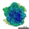
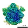
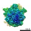
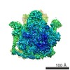
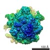
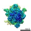
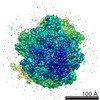
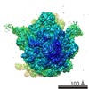
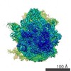
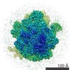
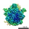
| Title | SecM-stalled ribosomes adopt an altered geometry at the peptidyltransferase center | |||||||||
 Map data Map data | SecM stalled ribosome | |||||||||
 Sample Sample |
| |||||||||
 Keywords Keywords | SecM-stalled RNC | |||||||||
| Biological species |  | |||||||||
| Method | single particle reconstruction / cryo EM / negative staining / Resolution: 5.6 Å | |||||||||
 Authors Authors | Bhushan S / Hoffman T / Seidelt B / Frauenfeld J / Mielke T / Berninghausen O / Wilson D / Beckmann R | |||||||||
 Citation Citation | Journal: PLoS Biol / Year: 2011 Title: SecM-stalled ribosomes adopt an altered geometry at the peptidyl transferase center. Authors: Shashi Bhushan / Thomas Hoffmann / Birgit Seidelt / Jens Frauenfeld / Thorsten Mielke / Otto Berninghausen / Daniel N Wilson / Roland Beckmann /  Abstract: As nascent polypeptide chains are synthesized, they pass through a tunnel in the large ribosomal subunit. Interaction between specific nascent chains and the ribosomal tunnel is used to induce ...As nascent polypeptide chains are synthesized, they pass through a tunnel in the large ribosomal subunit. Interaction between specific nascent chains and the ribosomal tunnel is used to induce translational stalling for the regulation of gene expression. One well-characterized example is the Escherichia coli SecM (secretion monitor) gene product, which induces stalling to up-regulate translation initiation of the downstream secA gene, which is needed for protein export. Although many of the key components of SecM and the ribosomal tunnel have been identified, understanding of the mechanism by which the peptidyl transferase center of the ribosome is inactivated has been lacking. Here we present a cryo-electron microscopy reconstruction of a SecM-stalled ribosome nascent chain complex at 5.6 Å. While no cascade of rRNA conformational changes is evident, this structure reveals the direct interaction between critical residues of SecM and the ribosomal tunnel. Moreover, a shift in the position of the tRNA-nascent peptide linkage of the SecM-tRNA provides a rationale for peptidyl transferase center silencing, conditional on the simultaneous presence of a Pro-tRNA(Pro) in the ribosomal A-site. These results suggest a distinct allosteric mechanism of regulating translational elongation by the SecM stalling peptide. | |||||||||
| History |
|
- Structure visualization
| Movie |
 Movie viewer Movie viewer |
|---|---|
| Structure viewer | EM map: SurfViewMolmilJmol/JSmol |
| Supplemental images |

- Downloads & links
-EMDB archive
| Map data | emd_1829.map.gz | 15.7 MB | EMDB map data format | |
|---|---|---|---|---|
| Header (meta data) | emd-1829-v30.xmlemd-1829.xml | 9.8 KB 9.8 KB | Display Display | EMDB header |
| Images | 1829.tif | 968.4 KB | ||
| Archive directory |  http://ftp.pdbj.org/pub/emdb/structures/EMD-1829ftp://ftp.pdbj.org/pub/emdb/structures/EMD-1829 http://ftp.pdbj.org/pub/emdb/structures/EMD-1829ftp://ftp.pdbj.org/pub/emdb/structures/EMD-1829 | HTTPS FTP |
-Validation report
| Summary document | emd_1829_validation.pdf.gz | 273.1 KB | Display | EMDB validaton report |
|---|---|---|---|---|
| Full document | emd_1829_full_validation.pdf.gz | 272.2 KB | Display | |
| Data in XML | emd_1829_validation.xml.gz | 6.7 KB | Display | |
| Arichive directory | https://ftp.pdbj.org/pub/emdb/validation_reports/EMD-1829ftp://ftp.pdbj.org/pub/emdb/validation_reports/EMD-1829 | HTTPS FTP |
-Related structure data
-Links
| EMDB pages | EMDB (EBI/PDBe) / EMDataResource |
|---|---|
| Related items in Molecule of the Month |

-Map
| File | Download / File: emd_1829.map.gz / Format: CCP4 / Size: 94.7 MB / Type: IMAGE STORED AS FLOATING POINT NUMBER (4 BYTES) | ||||||||||||||||||||||||||||||||||||||||||||||||||||||||||||||||||||
|---|---|---|---|---|---|---|---|---|---|---|---|---|---|---|---|---|---|---|---|---|---|---|---|---|---|---|---|---|---|---|---|---|---|---|---|---|---|---|---|---|---|---|---|---|---|---|---|---|---|---|---|---|---|---|---|---|---|---|---|---|---|---|---|---|---|---|---|---|---|
| Annotation | SecM stalled ribosome | ||||||||||||||||||||||||||||||||||||||||||||||||||||||||||||||||||||
| Voxel size | X=Y=Z: 1.237 Å | ||||||||||||||||||||||||||||||||||||||||||||||||||||||||||||||||||||
| Density |
| ||||||||||||||||||||||||||||||||||||||||||||||||||||||||||||||||||||
| Symmetry | Space group: 1 | ||||||||||||||||||||||||||||||||||||||||||||||||||||||||||||||||||||
| Details | EMDB XML:
CCP4 map header:
| ||||||||||||||||||||||||||||||||||||||||||||||||||||||||||||||||||||
-Supplemental data
- Sample components
-Entire : SecM-stalled RNC
| Entire | Name: SecM-stalled RNC |
|---|---|
| Components |
|
-Supramolecule #1000: SecM-stalled RNC
| Supramolecule | Name: SecM-stalled RNC / type: sample / ID: 1000 / Details: no / Number unique components: 1 |
|---|---|
| Molecular weight | Experimental: 2.3 MDa / Theoretical: 2.3 MDa |
-Supramolecule #1: 70S
| Supramolecule | Name: 70S / type: complex / ID: 1 / Name.synonym: 70S / Recombinant expression: No / Ribosome-details: ribosome-prokaryote: ALL |
|---|---|
| Source (natural) | Organism: |
| Molecular weight | Experimental: 2.3 MDa / Theoretical: 2.3 MDa |
-Experimental details
-Structure determination
| Method | negative staining, cryo EM |
|---|---|
 Processing Processing | single particle reconstruction |
| Aggregation state | particle |
-Sample preparation
| Buffer | pH: 7 Details: 20 mM Hepes Ph 7.0, 50 mM Pot Acetate, 6 mM Mag Acetate, 5 mM DTT, 500 ug/ml Chloroamphenicol, 0.05 % Nikkol, 0.5 % pill/ml, 0.1 U/ml RNAsin, 125 mM sucrose |
|---|---|
| Staining | Type: NEGATIVE / Details: native |
| Grid | Details: 2 nM carbon coated 200 mesh Cu grid |
| Vitrification | Cryogen name: ETHANE / Chamber humidity: 100 % / Instrument: OTHER / Details: Vitrification instrument: Vitrobot / Method: 10 seconds for blotting |
- Electron microscopy
| Microscope | FEI POLARA 300 |
|---|---|
| Specialist optics | Energy filter - Name: FEI |
| Image recording | Category: FILM / Film or detector model: KODAK SO-163 FILM / Digitization - Scanner: PRIMESCAN / Number real images: 400 / Average electron dose: 25 e/Å2 |
| Electron beam | Acceleration voltage: 300 kV / Electron source:  FIELD EMISSION GUN FIELD EMISSION GUN |
| Electron optics | Illumination mode: SPOT SCAN / Imaging mode: BRIGHT FIELD / Nominal defocus max: 4.0 µm / Nominal defocus min: 1.0 µm / Nominal magnification: 38900 |
| Sample stage | Specimen holder: Eucentric / Specimen holder model: GATAN LIQUID NITROGEN |
| Experimental equipment |  Model: Tecnai Polara / Image courtesy: FEI Company |
-Image processing
| Details | Processed using Spider |
|---|---|
| CTF correction | Details: CTFFIND on Micrographs |
| Final reconstruction | Applied symmetry - Point group: C1 (asymmetric) / Algorithm: OTHER / Resolution.type: BY AUTHOR / Resolution: 5.6 Å / Resolution method: FSC 0.5 CUT-OFF / Software - Name: Spider / Number images used: 544000 |
-Atomic model buiding 1
| Initial model | PDB ID:  2wwq |
|---|---|
| Software | Name: Chimera |
| Details | Fitted manually using Chimera and Coot |
| Refinement | Space: REAL / Protocol: RIGID BODY FIT |
-Atomic model buiding 2
| Initial model | PDB ID: 2wwl |
|---|---|
| Software | Name: Chimera |
| Details | Fitted manually using Chimera and Coot |
| Refinement | Space: REAL / Protocol: RIGID BODY FIT |
