Movie
Movie Controller

[English] 日本語
 Yorodumi
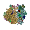
- EMDB-6485: Mechanisms of Ribosome Stalling by SecM at Multiple Elongation Steps -
+ Open data

- Basic information
| Entry | Database: EMDB / ID: EMD-6485 | |||||||||
|---|---|---|---|---|---|---|---|---|---|---|
| Title | Mechanisms of Ribosome Stalling by SecM at Multiple Elongation Steps | |||||||||
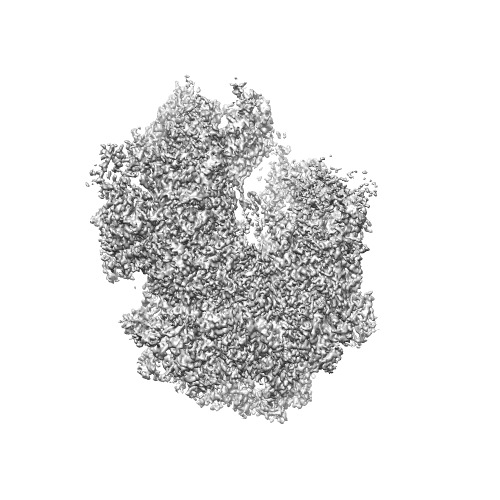
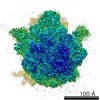
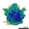
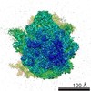
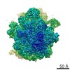
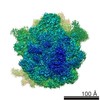
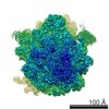
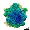
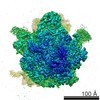
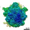
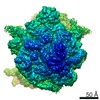
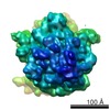
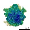
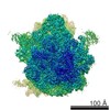
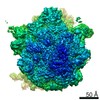
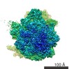
 Map data Map data | Cryo EM reconstruction of SecM-Pro-RNC ( ribosomal-nascent-peptide-complex) without 50S-tRNA mask | |||||||||
 Sample Sample |
| |||||||||
 Keywords Keywords |  electron microscopy / single particle analysis / ribosome stalling electron microscopy / single particle analysis / ribosome stalling | |||||||||
| Function / homology |  Function and homology information Function and homology informationouter membrane protein complex / monoatomic ion transmembrane transporter activity / detection of virus / outer membrane / porin activity / pore complex / mRNA base-pairing translational repressor activity / ornithine decarboxylase inhibitor activity / misfolded RNA binding / transcription antitermination factor activity, RNA binding ...outer membrane protein complex / monoatomic ion transmembrane transporter activity / detection of virus / outer membrane / porin activity / pore complex / mRNA base-pairing translational repressor activity / ornithine decarboxylase inhibitor activity / misfolded RNA binding / transcription antitermination factor activity, RNA binding / Group I intron splicing / RNA folding / transcriptional attenuation / endoribonuclease inhibitor activity / RNA-binding transcription regulator activity / positive regulation of ribosome biogenesis / negative regulation of cytoplasmic translation / monoatomic ion transport / DnaA-L2 complex / four-way junction DNA binding / negative regulation of translational initiation / translation repressor activity / negative regulation of DNA-templated DNA replication initiation / regulation of mRNA stability / ribosome assembly / mRNA regulatory element binding translation repressor activity / response to reactive oxygen species / assembly of large subunit precursor of preribosome / positive regulation of RNA splicing / DNA endonuclease activity / transcription elongation factor complex / cytosolic ribosome assembly / regulation of DNA-templated transcription elongation / transcription antitermination / regulation of cell growth / maintenance of translational fidelity / DNA-templated transcription termination / cell outer membrane / response to radiation / mRNA 5'-UTR binding / ribosomal small subunit biogenesis / ribosomal large subunit assembly / small ribosomal subunit rRNA binding / ribosomal small subunit assembly / cytosolic small ribosomal subunit / large ribosomal subunit rRNA binding / ribosome binding / large ribosomal subunit / ribosome biogenesis / regulation of translation / outer membrane-bounded periplasmic space / cytoplasmic translation / small ribosomal subunit / 5S rRNA binding / cytosolic large ribosomal subunit / transferase activity / tRNA binding / negative regulation of translation / rRNA binding / molecular adaptor activity / ribosome / structural constituent of ribosome / symbiont entry into host cell / translation / response to antibiotic / mRNA binding / negative regulation of DNA-templated transcription / DNA damage response / DNA binding / RNA binding / zinc ion binding / membrane / identical protein binding / cytosol / cytoplasmSimilarity search - Function | |||||||||
| Biological species |  Escherichia coli (E. coli) Escherichia coli (E. coli) | |||||||||
| Method | single particle reconstruction / cryo EM / Resolution: 3.32 Å | |||||||||
 Authors Authors | Zhang J / Pan XJ / Yan KG / Sun S / Gao N / Sui SF | |||||||||
 Citation Citation | Journal: Elife / Year: 2015 Title: Mechanisms of ribosome stalling by SecM at multiple elongation steps. Authors: Jun Zhang / Xijiang Pan / Kaige Yan / Shan Sun / Ning Gao / Sen-Fang Sui /  Abstract: Regulation of translating ribosomes is a major component of gene expression control network. In Escherichia coli, ribosome stalling by the C-terminal arrest sequence of SecM regulates the SecA- ...Regulation of translating ribosomes is a major component of gene expression control network. In Escherichia coli, ribosome stalling by the C-terminal arrest sequence of SecM regulates the SecA-dependent secretion pathway. Previous studies reported many residues of SecM peptide and ribosome exit tunnel are critical for stalling. However, the underlying molecular mechanism is still not clear at the atomic level. Here, we present two cryo-EM structures of the SecM-stalled ribosomes at 3.3-3.7 Å resolution, which reveal two different stalling mechanisms at distinct elongation steps of the translation cycle: one is due to the inactivation of ribosomal peptidyl-transferase center which inhibits peptide bond formation with the incoming prolyl-tRNA; the other is the prolonged residence of the peptidyl-RNA at the hybrid A/P site which inhibits the full-scale tRNA translocation. These results demonstrate an elegant control of translation cycle by regulatory peptides through a continuous, dynamic reshaping of the functional center of the ribosome. | |||||||||
| History |
|
- Structure visualization
| Movie |
Movie viewer |
|---|---|
| Structure viewer | EM map: SurfViewMolmilJmol/JSmol |
| Supplemental images |
- Downloads & links
-EMDB archive
| Map data | emd_6485.map.gz | 117.1 MB | EMDB map data format | |
|---|---|---|---|---|
| Header (meta data) | emd-6485-v30.xmlemd-6485.xml | 13.8 KB 13.8 KB | Display Display | EMDB header |
| FSC (resolution estimation) | emd_6485_fsc.xml | 11.3 KB | Display | FSC data file |
| Images | emd_6485.tif | 130 KB | ||
| Archive directory |  http://ftp.pdbj.org/pub/emdb/structures/EMD-6485ftp://ftp.pdbj.org/pub/emdb/structures/EMD-6485 http://ftp.pdbj.org/pub/emdb/structures/EMD-6485ftp://ftp.pdbj.org/pub/emdb/structures/EMD-6485 | HTTPS FTP |
-Related structure data

-Links
| EMDB pages | EMDB (EBI/PDBe) / EMDataResource |
|---|---|
| Related items in Molecule of the Month |


-Map
| File | Download / File: emd_6485.map.gz / Format: CCP4 / Size: 122.1 MB / Type: IMAGE STORED AS FLOATING POINT NUMBER (4 BYTES) | ||||||||||||||||||||||||||||||||||||||||||||||||||||||||||||
|---|---|---|---|---|---|---|---|---|---|---|---|---|---|---|---|---|---|---|---|---|---|---|---|---|---|---|---|---|---|---|---|---|---|---|---|---|---|---|---|---|---|---|---|---|---|---|---|---|---|---|---|---|---|---|---|---|---|---|---|---|---|
| Annotation | Cryo EM reconstruction of SecM-Pro-RNC ( ribosomal-nascent-peptide-complex) without 50S-tRNA mask | ||||||||||||||||||||||||||||||||||||||||||||||||||||||||||||
| Voxel size | X=Y=Z: 1.32 Å | ||||||||||||||||||||||||||||||||||||||||||||||||||||||||||||
| Density |
| ||||||||||||||||||||||||||||||||||||||||||||||||||||||||||||
| Symmetry | Space group: 1 | ||||||||||||||||||||||||||||||||||||||||||||||||||||||||||||
| Details | EMDB XML:
CCP4 map header:
| ||||||||||||||||||||||||||||||||||||||||||||||||||||||||||||
-Supplemental data
- Sample components
-Entire : SecM-Pro-RNC(ribosome-nascent-peptide-complex) complex without mask
| Entire | Name: SecM-Pro-RNC(ribosome-nascent-peptide-complex) complex without mask |
|---|---|
| Components |
|
-Supramolecule #1000: SecM-Pro-RNC(ribosome-nascent-peptide-complex) complex without mask
| Supramolecule | Name: SecM-Pro-RNC(ribosome-nascent-peptide-complex) complex without mask type: sample / ID: 1000 / Number unique components: 1 |
|---|---|
| Molecular weight | Theoretical: 2.3 MDa |
-Supramolecule #1: SecM-Pro-RNC without mask
| Supramolecule | Name: SecM-Pro-RNC without mask / type: complex / ID: 1 / Name.synonym: SecM-Pro-stalled RNC without mask / Recombinant expression: Yes / Ribosome-details: ribosome-prokaryote: ALL |
|---|---|
| Source (natural) | Organism: Escherichia coli (E. coli) |
| Recombinant expression | Organism: Escherichia coli (E. coli) / Recombinant strain: Escherichia coli MG1655 / Recombinant plasmid: pET21 |
| Molecular weight | Theoretical: 2.3 MDa |
-Experimental details
-Structure determination
| Method | cryo EM |
|---|---|
 Processing Processing | single particle reconstruction |
| Aggregation state | particle |
-Sample preparation
| Buffer | pH: 7 Details: 20mM HEPES, 50mM KOAc, 6mM Mg[OAc]2, 1mM DTT, 500 ug/ml chloramphenicol,0.05% Nikkol,0.5% pill/ml Complete EDTA-free Protease inhibitor cocktail,0.1 U/ml RNasin and 125mM sucrose |
|---|---|
| Vitrification | Cryogen name: ETHANE / Chamber humidity: 100 % / Chamber temperature: 277.15 K / Instrument: FEI VITROBOT MARK IV / Method: Blot for 1.5 seconds before plunging |
- Electron microscopy #1
| Microscope | FEI TITAN KRIOS |
|---|---|
| Electron beam | Acceleration voltage: 300 kV / Electron source: FIELD EMISSION GUN |
| Electron optics | Calibrated magnification: 37878 / Illumination mode: FLOOD BEAM / Imaging mode: BRIGHT FIELDBright-field microscopy / Cs: 2.7 mm / Nominal defocus max: 3.5 µm / Nominal defocus min: 1.0 µm / Nominal magnification: 22500 |
| Sample stage | Specimen holder model: FEI TITAN KRIOS AUTOGRID HOLDER |
| Microscopy ID | 1 |
| Date | May 8, 2014 |
| Image recording | Category: CCD / Film or detector model: GATAN K2 (4k x 4k) / Digitization - Sampling interval: 4 µm / Number real images: 3908 / Average electron dose: 16 e/Å2 Details: Every image is the average of 14 frames recorded by the direct electron detector |
| Experimental equipment |  Model: Titan Krios / Image courtesy: FEI Company |
-Electron microscopy #2
| Microscope | FEI TITAN KRIOS |
|---|---|
| Electron beam | Acceleration voltage: 300 kV / Electron source: FIELD EMISSION GUN |
| Electron optics | Calibrated magnification: 37878 / Illumination mode: FLOOD BEAM / Imaging mode: BRIGHT FIELDBright-field microscopy / Cs: 2.7 mm / Nominal defocus max: 3.5 µm / Nominal defocus min: 1.0 µm / Nominal magnification: 22500 |
| Sample stage | Specimen holder model: FEI TITAN KRIOS AUTOGRID HOLDER |
| Microscopy ID | 2 |
| Date | Jun 16, 2014 |
| Image recording | Category: CCD / Film or detector model: GATAN K2 (4k x 4k) / Digitization - Sampling interval: 4 µm / Number real images: 3908 / Average electron dose: 16 e/Å2 Details: Every image is the average of 14 frames recorded by the direct electron detector |
| Experimental equipment | Model: Titan Krios / Image courtesy: FEI Company |
-Electron microscopy #3
| Microscope | FEI TITAN KRIOS |
|---|---|
| Electron beam | Acceleration voltage: 300 kV / Electron source: FIELD EMISSION GUN |
| Electron optics | Calibrated magnification: 37878 / Illumination mode: FLOOD BEAM / Imaging mode: BRIGHT FIELDBright-field microscopy / Cs: 2.7 mm / Nominal defocus max: 3.5 µm / Nominal defocus min: 1.0 µm / Nominal magnification: 22500 |
| Sample stage | Specimen holder model: FEI TITAN KRIOS AUTOGRID HOLDER |
| Microscopy ID | 3 |
| Date | Aug 30, 2014 |
| Image recording | Category: CCD / Film or detector model: GATAN K2 (4k x 4k) / Digitization - Sampling interval: 4 µm / Number real images: 3908 / Average electron dose: 16 e/Å2 Details: Every image is the average of 14 frames recorded by the direct electron detector |
| Experimental equipment | Model: Titan Krios / Image courtesy: FEI Company |
-Image processing
| CTF correction | Details: CTFFIND |
|---|---|
| Final reconstruction | Algorithm: OTHER / Resolution.type: BY AUTHOR / Resolution: 3.32 Å / Resolution method: OTHER / Software - Name: RELION / Number images used: 60354 |
| FSC plot (resolution estimation) | 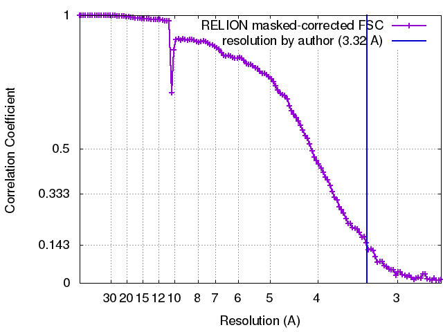 |
-Atomic model buiding 1
| Initial model | PDB ID: Chain - #0 - Chain ID: A / Chain - #1 - Chain ID: B |
|---|---|
| Software | Name: Chimera, EMFit, Coot |
| Refinement | Space: RECIPROCAL / Protocol: FLEXIBLE FIT |