 Movie
Movie Controller

[English] 日本語
 Yorodumi
- PDB-3vmx: Crystal Structure of a parallel coiled-coil dimerization domain f... -
+ Open data

- Basic information
| Entry | Database: PDB / ID: 3vmx | ||||||
|---|---|---|---|---|---|---|---|
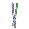
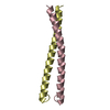
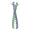
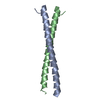
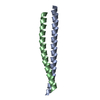
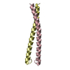
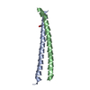
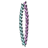
| Title | Crystal Structure of a parallel coiled-coil dimerization domain from the voltage-gated proton channel | ||||||
 Components Components | Voltage-gated hydrogen channel 1 | ||||||
 Keywords Keywords |  MEMBRANE PROTEIN / COILED-COIL / ION CHANNEL / ION TRANSPORT MEMBRANE PROTEIN / COILED-COIL / ION CHANNEL / ION TRANSPORT | ||||||
| Function / homology |  Function and homology informationvoltage-gated proton channel activity / Sperm Motility And Taxes / ROS and RNS production in phagocytes / cellular response to pH / voltage-gated monoatomic cation channel activity / response to pH / regulation of reactive oxygen species biosynthetic process / cellular response to zinc ion / monoatomic ion channel complex / response to zinc ion ...voltage-gated proton channel activity / Sperm Motility And Taxes / ROS and RNS production in phagocytes / cellular response to pH / voltage-gated monoatomic cation channel activity / response to pH / regulation of reactive oxygen species biosynthetic process / cellular response to zinc ion / monoatomic ion channel complex / response to zinc ion / proton transmembrane transport / Neutrophil degranulation / positive regulation of superoxide anion generation / regulation of intracellular pH / phagocytic vesicle membrane / apical plasma membrane / membrane / identical protein binding / plasma membrane Function and homology informationvoltage-gated proton channel activity / Sperm Motility And Taxes / ROS and RNS production in phagocytes / cellular response to pH / voltage-gated monoatomic cation channel activity / response to pH / regulation of reactive oxygen species biosynthetic process / cellular response to zinc ion / monoatomic ion channel complex / response to zinc ion ...voltage-gated proton channel activity / Sperm Motility And Taxes / ROS and RNS production in phagocytes / cellular response to pH / voltage-gated monoatomic cation channel activity / response to pH / regulation of reactive oxygen species biosynthetic process / cellular response to zinc ion / monoatomic ion channel complex / response to zinc ion / proton transmembrane transport / Neutrophil degranulation / positive regulation of superoxide anion generation / regulation of intracellular pH / phagocytic vesicle membrane / apical plasma membrane / membrane / identical protein binding / plasma membraneSimilarity search - Function | ||||||
| Biological species |  Mus musculus (house mouse) Mus musculus (house mouse) | ||||||
| Method | X-RAY DIFFRACTION / SYNCHROTRON / MOLECULAR REPLACEMENT / Resolution: 1.45 Å | ||||||
 Authors Authors | Fujiwara, Y. / Takeshita, K. / Kobayashi, M. / Okamura, Y. / Nakagawa, A. | ||||||
 Citation Citation | Journal: Nat Commun / Year: 2012 Title: The cytoplasmic coiled-coil mediates cooperative gating temperature sensitivity in the voltage-gated H(+) channel Hv1 Authors: Fujiwara, Y. / Kurokawa, T. / Takeshita, K. / Kobayashi, M. / Okochi, Y. / Nakagawa, A. / Okamura, Y. | ||||||
| History |
|
- Structure visualization
| Structure viewer | Molecule: MolmilJmol/JSmol |
|---|
- Downloads & links
-Download
| PDBx/mmCIF format | 3vmx.cif.gz | 96.3 KB | Display | PDBx/mmCIF format |
|---|---|---|---|---|
| PDB format | pdb3vmx.ent.gz | 78 KB | Display | PDB format |
| PDBx/mmJSON format | 3vmx.json.gz | Tree view | PDBx/mmJSON format | |
| Others |  Other downloads Other downloads |
-Validation report
| Arichive directory | https://data.pdbj.org/pub/pdb/validation_reports/vm/3vmxftp://data.pdbj.org/pub/pdb/validation_reports/vm/3vmx | HTTPS FTP |
|---|
-Related structure data
| Related structure data | |
|---|---|
| Similar structure data |


-Links
 PDBj
PDBj


- Assembly
| Deposited unit | 
| ||||||||
|---|---|---|---|---|---|---|---|---|---|
| 1 | 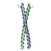
| ||||||||
| 2 | 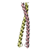
| ||||||||
| Unit cell |
|
-Components
| #1: Protein/peptide | Mass: 5598.501 Da / Num. of mol.: 4 / Fragment: C-TERMINAL DOMAIN (UNP RESIDUES 220-269) Source method: isolated from a genetically manipulated source Source: (gene. exp.) Mus musculus (house mouse) / Gene: Bts, Hvcn1, Vsop / Plasmid: PET28HMT / Production host:  ESCHERICHIA COLI (E. coli) / Strain (production host): BL21PLYSS / References: UniProt: Q3U2S8 ESCHERICHIA COLI (E. coli) / Strain (production host): BL21PLYSS / References: UniProt: Q3U2S8#2: Water | ChemComp-HOH / | Water Mass: 18.015 Da / Num. of mol.: 222 / Source method: isolated from a natural source / Formula: H2O Mass: 18.015 Da / Num. of mol.: 222 / Source method: isolated from a natural source / Formula: H2O |
|---|
-Experimental details
-Experiment
| Experiment | Method: X-RAY DIFFRACTION / Number of used crystals: 1 |
|---|
- Sample preparation
| Crystal | Density Matthews: 1.97 Å3/Da / Density % sol: 37.62 % |
|---|---|
| Crystal grow | Temperature: 283 K / Method: vapor diffusion, sitting drop / pH: 7 Details: 2% V/V TACSIMATE (PH 7.0), 0.1M IMIDAZOLE (PH 7.0), 8% W/V PEG 3350, 5% V/V 2-PROPANOL (PEGRX-2 # 26, HAMPTOM), VAPOR DIFFUSION, SITTING DROP, TEMPERATURE 283K |
-Data collection
| Diffraction | Mean temperature: 200 K |
|---|---|
| Diffraction source | Source: SYNCHROTRON / Site: SPring-8  / Beamline: BL44XU / Wavelength: 0.9 / Beamline: BL44XU / Wavelength: 0.9 |
| Detector | Type: RAYONIX MX225HE / Detector: CCD / Date: Nov 22, 2009 |
| Radiation | Protocol: SINGLE WAVELENGTH / Monochromatic (M) / Laue (L): M / Scattering type: x-ray |
| Radiation wavelength | Wavelength: 0.9 Å / Relative weight: 1 |
| Reflection | Resolution: 1.45→50 Å / Num. obs: 232980 / % possible obs: 99.7 % / Observed criterion σ(I): -3 / Redundancy: 7.2 % / Rmerge(I) obs: 0.056 / Rsym value: 0.056 / Net I/σ(I): 43.972 |
| Reflection shell | Resolution: 1.45→1.48 Å / Redundancy: 7.3 % / Rmerge(I) obs: 0.44 / Mean I/σ(I) obs: 4.741 / Rsym value: 0.44 / % possible all: 100 |
- Processing
| Software |
| ||||||||||||||||||||||||||||||||||||||||||||||||||||||||||||||||||||||||||||||||||||||||||||||||||||||||||||||||||||||||||||||||||||||||||||||||||||||||||||||||||||||||||
|---|---|---|---|---|---|---|---|---|---|---|---|---|---|---|---|---|---|---|---|---|---|---|---|---|---|---|---|---|---|---|---|---|---|---|---|---|---|---|---|---|---|---|---|---|---|---|---|---|---|---|---|---|---|---|---|---|---|---|---|---|---|---|---|---|---|---|---|---|---|---|---|---|---|---|---|---|---|---|---|---|---|---|---|---|---|---|---|---|---|---|---|---|---|---|---|---|---|---|---|---|---|---|---|---|---|---|---|---|---|---|---|---|---|---|---|---|---|---|---|---|---|---|---|---|---|---|---|---|---|---|---|---|---|---|---|---|---|---|---|---|---|---|---|---|---|---|---|---|---|---|---|---|---|---|---|---|---|---|---|---|---|---|---|---|---|---|---|---|---|---|---|
| Refinement | Method to determine structure: MOLECULAR REPLACEMENT / Resolution: 1.45→45.01 Å / Cor.coef. Fo:Fc: 0.961 / Cor.coef. Fo:Fc free: 0.949 / SU B: 2.784 / SU ML: 0.049 / Cross valid method: THROUGHOUT / σ(F): 0 / ESU R Free: 0.08 / Stereochemistry target values: MAXIMUM LIKELIHOOD Details: HYDROGENS HAVE BEEN ADDED IN THE RIDING POSITIONS U VALUES
| ||||||||||||||||||||||||||||||||||||||||||||||||||||||||||||||||||||||||||||||||||||||||||||||||||||||||||||||||||||||||||||||||||||||||||||||||||||||||||||||||||||||||||
| Solvent computation | Ion probe radii: 0.8 Å / Shrinkage radii: 0.8 Å / VDW probe radii: 1.4 Å / Solvent model: MASK | ||||||||||||||||||||||||||||||||||||||||||||||||||||||||||||||||||||||||||||||||||||||||||||||||||||||||||||||||||||||||||||||||||||||||||||||||||||||||||||||||||||||||||
| Displacement parameters | Biso mean: 19.72 Å2
| ||||||||||||||||||||||||||||||||||||||||||||||||||||||||||||||||||||||||||||||||||||||||||||||||||||||||||||||||||||||||||||||||||||||||||||||||||||||||||||||||||||||||||
| Refinement step | Cycle: LAST / Resolution: 1.45→45.01 Å
| ||||||||||||||||||||||||||||||||||||||||||||||||||||||||||||||||||||||||||||||||||||||||||||||||||||||||||||||||||||||||||||||||||||||||||||||||||||||||||||||||||||||||||
| Refine LS restraints |
| ||||||||||||||||||||||||||||||||||||||||||||||||||||||||||||||||||||||||||||||||||||||||||||||||||||||||||||||||||||||||||||||||||||||||||||||||||||||||||||||||||||||||||
| LS refinement shell | Resolution: 1.45→1.49 Å / Total num. of bins used: 20
| ||||||||||||||||||||||||||||||||||||||||||||||||||||||||||||||||||||||||||||||||||||||||||||||||||||||||||||||||||||||||||||||||||||||||||||||||||||||||||||||||||||||||||
| Refinement TLS params. | Method: refined / Refine-ID: X-RAY DIFFRACTION
| ||||||||||||||||||||||||||||||||||||||||||||||||||||||||||||||||||||||||||||||||||||||||||||||||||||||||||||||||||||||||||||||||||||||||||||||||||||||||||||||||||||||||||
| Refinement TLS group |
|
