Movie
Movie Controller

[English] 日本語
 Yorodumi
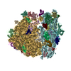
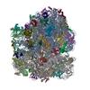
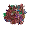
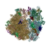
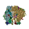
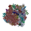
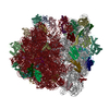
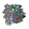
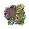
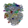
- PDB-3jbv: Mechanisms of Ribosome Stalling by SecM at Multiple Elongation Steps -
+ Open data

- Basic information
| Entry | Database: PDB / ID: 3jbv | ||||||
|---|---|---|---|---|---|---|---|
| Title | Mechanisms of Ribosome Stalling by SecM at Multiple Elongation Steps | ||||||
 Components Components |
| ||||||
 Keywords Keywords | RIBOSOME / single particle analysis / ribosome stalling | ||||||
| Function / homology |  Function and homology information Function and homology informationstringent response / ornithine decarboxylase inhibitor activity / transcription antitermination factor activity, RNA binding / misfolded RNA binding / Group I intron splicing / RNA folding / transcriptional attenuation / endoribonuclease inhibitor activity / RNA-binding transcription regulator activity / positive regulation of ribosome biogenesis ...stringent response / ornithine decarboxylase inhibitor activity / transcription antitermination factor activity, RNA binding / misfolded RNA binding / Group I intron splicing / RNA folding / transcriptional attenuation / endoribonuclease inhibitor activity / RNA-binding transcription regulator activity / positive regulation of ribosome biogenesis / negative regulation of cytoplasmic translation / translation regulator activity / four-way junction DNA binding / translational termination / DnaA-L2 complex / translation repressor activity / negative regulation of translational initiation / negative regulation of DNA-templated DNA replication initiation / regulation of mRNA stability / mRNA regulatory element binding translation repressor activity / ribosome assembly / positive regulation of RNA splicing / assembly of large subunit precursor of preribosome / transcription elongation factor complex / cytosolic ribosome assembly / regulation of DNA-templated transcription elongation / DNA endonuclease activity / ribosomal large subunit assembly / transcription antitermination / response to reactive oxygen species / regulation of cell growth / DNA-templated transcription termination / maintenance of translational fidelity / response to radiation / mRNA 5'-UTR binding / large ribosomal subunit / ribosome biogenesis / ribosome binding / regulation of translation / ribosomal small subunit biogenesis / ribosomal small subunit assembly / small ribosomal subunit / small ribosomal subunit rRNA binding / transferase activity / 5S rRNA binding / large ribosomal subunit rRNA binding / cytosolic small ribosomal subunit / cytosolic large ribosomal subunit / cytoplasmic translation / periplasmic space / tRNA binding / molecular adaptor activity / rRNA binding / negative regulation of translation / ribosome / structural constituent of ribosome / translation / response to antibiotic / negative regulation of DNA-templated transcription / mRNA binding / DNA binding / RNA binding / zinc ion binding / membrane / cytoplasm / cytosol Similarity search - Function | ||||||
| Biological species |  | ||||||
| Method | ELECTRON MICROSCOPY / single particle reconstruction / cryo EM / Resolution: 3.32 Å | ||||||
 Authors Authors | Zhang, J. / Pan, X.J. / Yan, K.G. / Sun, S. / Gao, N. / Sui, S.F. | ||||||
 Citation Citation | Journal: Elife / Year: 2015 Title: Mechanisms of ribosome stalling by SecM at multiple elongation steps. Authors: Jun Zhang / Xijiang Pan / Kaige Yan / Shan Sun / Ning Gao / Sen-Fang Sui /  Abstract: Regulation of translating ribosomes is a major component of gene expression control network. In Escherichia coli, ribosome stalling by the C-terminal arrest sequence of SecM regulates the SecA- ...Regulation of translating ribosomes is a major component of gene expression control network. In Escherichia coli, ribosome stalling by the C-terminal arrest sequence of SecM regulates the SecA-dependent secretion pathway. Previous studies reported many residues of SecM peptide and ribosome exit tunnel are critical for stalling. However, the underlying molecular mechanism is still not clear at the atomic level. Here, we present two cryo-EM structures of the SecM-stalled ribosomes at 3.3-3.7 Å resolution, which reveal two different stalling mechanisms at distinct elongation steps of the translation cycle: one is due to the inactivation of ribosomal peptidyl-transferase center which inhibits peptide bond formation with the incoming prolyl-tRNA; the other is the prolonged residence of the peptidyl-RNA at the hybrid A/P site which inhibits the full-scale tRNA translocation. These results demonstrate an elegant control of translation cycle by regulatory peptides through a continuous, dynamic reshaping of the functional center of the ribosome. | ||||||
| History |
|
- Structure visualization
| Movie |
Movie viewer |
|---|---|
| Structure viewer | Molecule: MolmilJmol/JSmol |
- Downloads & links
-Download
| PDBx/mmCIF format | 3jbv.cif.gz | 3.4 MB | Display | PDBx/mmCIF format |
|---|---|---|---|---|
| PDB format | pdb3jbv.ent.gz | Display | PDB format | |
| PDBx/mmJSON format | 3jbv.json.gz | Tree view | PDBx/mmJSON format | |
| Others |  Other downloads Other downloads |
-Validation report
| Summary document | 3jbv_validation.pdf.gz | 1.6 MB | Display | wwPDB validaton report |
|---|---|---|---|---|
| Full document | 3jbv_full_validation.pdf.gz | 1.8 MB | Display | |
| Data in XML | 3jbv_validation.xml.gz | 209.3 KB | Display | |
| Data in CIF | 3jbv_validation.cif.gz | 364.9 KB | Display | |
| Arichive directory | https://data.pdbj.org/pub/pdb/validation_reports/jb/3jbvftp://data.pdbj.org/pub/pdb/validation_reports/jb/3jbv | HTTPS FTP |
-Related structure data
| Related structure data | 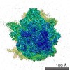 6486MC 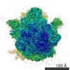 6483C 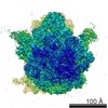 6484C 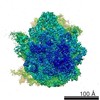 6485C 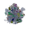 3jbuC C: citing same article ( M: map data used to model this data |
|---|---|
| Similar structure data |

-Links
 PDBj
PDBj


- Assembly
| Deposited unit | 
|
|---|---|
| 1 |
|
-Components
-RNA chain , 6 types, 6 molecules AVWXab
| #1: RNA chain | Mass: 499690.031 Da / Num. of mol.: 1 / Source method: isolated from a natural source / Source: (natural) |
|---|---|
| #22: RNA chain | Mass: 24501.539 Da / Num. of mol.: 1 / Source method: isolated from a natural source / Source: (natural) |
| #23: RNA chain | Mass: 24186.365 Da / Num. of mol.: 1 / Source method: isolated from a natural source / Source: (natural) |
| #24: RNA chain | Mass: 3442.106 Da / Num. of mol.: 1 / Source method: isolated from a natural source / Source: (natural) |
| #33: RNA chain | Mass: 38790.090 Da / Num. of mol.: 1 / Source method: isolated from a natural source / Source: (natural) |
| #34: RNA chain | Mass: 941612.375 Da / Num. of mol.: 1 / Source method: isolated from a natural source / Source: (natural) |
-30S ribosomal protein ... , 20 types, 20 molecules BCDEFGHIJKLMNOPQRSTU
| #2: Protein | Mass: 26781.670 Da / Num. of mol.: 1 / Source method: isolated from a natural source / Source: (natural) |
|---|---|
| #3: Protein | Mass: 26031.316 Da / Num. of mol.: 1 / Source method: isolated from a natural source / Source: (natural) |
| #4: Protein | Mass: 23514.199 Da / Num. of mol.: 1 / Source method: isolated from a natural source / Source: (natural) |
| #5: Protein | Mass: 17629.398 Da / Num. of mol.: 1 / Source method: isolated from a natural source / Source: (natural) |
| #6: Protein | Mass: 15197.032 Da / Num. of mol.: 1 / Source method: isolated from a natural source / Source: (natural) |
| #7: Protein | Mass: 17637.445 Da / Num. of mol.: 1 / Source method: isolated from a natural source / Source: (natural) |
| #8: Protein | Mass: 14146.557 Da / Num. of mol.: 1 / Source method: isolated from a natural source / Source: (natural) |
| #9: Protein | Mass: 14886.270 Da / Num. of mol.: 1 / Source method: isolated from a natural source / Source: (natural) |
| #10: Protein | Mass: 11755.597 Da / Num. of mol.: 1 / Source method: isolated from a natural source / Source: (natural) |
| #11: Protein | Mass: 13870.975 Da / Num. of mol.: 1 / Fragment: UNP residues 1-131 / Source method: isolated from a natural source / Source: (natural) |
| #12: Protein | Mass: 13768.157 Da / Num. of mol.: 1 / Fragment: UNP residues 1-156 / Source method: isolated from a natural source / Source: (natural) |
| #13: Protein | Mass: 13128.467 Da / Num. of mol.: 1 / Fragment: UNP residues 1-120 / Source method: isolated from a natural source / Source: (natural) |
| #14: Protein | Mass: 11606.560 Da / Num. of mol.: 1 / Source method: isolated from a natural source / Source: (natural) |
| #15: Protein | Mass: 10319.882 Da / Num. of mol.: 1 / Source method: isolated from a natural source / Source: (natural) |
| #16: Protein | Mass: 9207.572 Da / Num. of mol.: 1 / Source method: isolated from a natural source / Source: (natural) |
| #17: Protein | Mass: 9724.491 Da / Num. of mol.: 1 / Source method: isolated from a natural source / Source: (natural) |
| #18: Protein | Mass: 9005.472 Da / Num. of mol.: 1 / Source method: isolated from a natural source / Source: (natural) |
| #19: Protein | Mass: 10455.355 Da / Num. of mol.: 1 / Source method: isolated from a natural source / Source: (natural) |
| #20: Protein | Mass: 9708.464 Da / Num. of mol.: 1 / Source method: isolated from a natural source / Source: (natural) |
| #21: Protein | Mass: 8524.039 Da / Num. of mol.: 1 / Source method: isolated from a natural source / Source: (natural) |
+50S ribosomal protein ... , 29 types, 29 molecules 01234678cidefghjklmnopqrstuwy
-Protein/peptide / Non-polymers , 2 types, 2 molecules z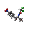
| #56: Protein/peptide | Mass: 3002.334 Da / Num. of mol.: 1 / Source method: isolated from a natural source / Source: (natural) |
|---|---|
| #57: Chemical | ChemComp-CLM /  Mass: 323.129 Da / Num. of mol.: 1 / Source method: obtained synthetically / Formula: C11H12Cl2N2O5 / Comment: antibiotic*YM Mass: 323.129 Da / Num. of mol.: 1 / Source method: obtained synthetically / Formula: C11H12Cl2N2O5 / Comment: antibiotic*YM |
-Details
| Sequence details | THE SEQUENCE OF THE CHAIN Z (ENTITY 56, UNCAPITALIZED Z) WAS NOT AVAILABLE AT THE UNIPROT ...THE SEQUENCE OF THE CHAIN Z (ENTITY 56, UNCAPITALI |
|---|
-Experimental details
-Experiment
| Experiment | Method: ELECTRON MICROSCOPY |
|---|---|
| EM experiment | Aggregation state: PARTICLE / 3D reconstruction method: single particle reconstruction |
- Sample preparation
| Component | Name: SecM-Pro-RNC / Type: RIBOSOME |
|---|---|
| Buffer solution | Name: 20mM HEPES, 50mM KOAc, 6mM Mg(OAc)2, 1mM DTT, 500 ug/ml chloramphenicol,0.05% Nikkol,0.5% pill/ml Complete EDTA-free Protease inhibitor cocktail,0.1 U/ml RNasin and 125mM sucrose pH: 7 Details: 20mM HEPES, 50mM KOAc, 6mM Mg(OAc)2, 1mM DTT, 500 ug/ml chloramphenicol,0.05% Nikkol,0.5% pill/ml Complete EDTA-free Protease inhibitor cocktail,0.1 U/ml RNasin and 125mM sucrose |
| Specimen | Embedding applied: NO / Shadowing applied: NO / Staining applied: NO / Vitrification applied: YES |
| Specimen support | Details: Sample are made by Cryo-EM on the holy carbon |
| Vitrification | Instrument: FEI VITROBOT MARK IV / Cryogen name: ETHANE |
- Electron microscopy imaging
| Experimental equipment |  Model: Titan Krios / Image courtesy: FEI Company |
|---|---|
| Microscopy | Model: FEI TITAN KRIOS / Date: May 8, 2014 |
| Electron gun | Electron source:  FIELD EMISSION GUN / Accelerating voltage: 300 kV / Illumination mode: FLOOD BEAM FIELD EMISSION GUN / Accelerating voltage: 300 kV / Illumination mode: FLOOD BEAM |
| Electron lens | Mode: BRIGHT FIELD / Nominal magnification: 22500 X / Calibrated magnification: 37878 X / Nominal defocus max: 1000 nm / Nominal defocus min: 3500 nm / Cs: 2.7 mm |
| Image recording | Electron dose: 16 e/Å2 / Film or detector model: GATAN K2 SUMMIT (4k x 4k) |
- Processing
| EM software |
| |||||||||||||||
|---|---|---|---|---|---|---|---|---|---|---|---|---|---|---|---|---|
| CTF correction | Details: CTFFIND | |||||||||||||||
| Symmetry | Point symmetry: C1 (asymmetric) | |||||||||||||||
| 3D reconstruction | Method: REFERENCE-BASED RECONSTRUCTION / Resolution: 3.32 Å / Num. of particles: 60354 / Nominal pixel size: 1.32 Å / Actual pixel size: 1.31 Å / Symmetry type: POINT | |||||||||||||||
| Atomic model building | Protocol: FLEXIBLE FIT / Space: RECIPROCAL / Details: METHOD--FLEXIBLE | |||||||||||||||
| Atomic model building | PDB-ID: 4V7T Accession code: 4V7T / Source name: PDB / Type: experimental model | |||||||||||||||
| Refinement step | Cycle: LAST
|