Movie
Movie Controller

[English] 日本語
 Yorodumi
- PDB-3j0g: Homology model of E3 protein of Venezuelan Equine Encephalitis Vi... -
+ Open data

- Basic information
| Entry | Database: PDB / ID: 3j0g | ||||||
|---|---|---|---|---|---|---|---|
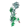
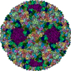
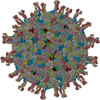
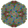
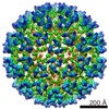
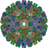
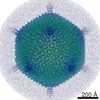
| Title | Homology model of E3 protein of Venezuelan Equine Encephalitis Virus TC-83 strain fitted with a cryo-EM map | ||||||
 Components Components | E3 protein | ||||||
 Keywords Keywords | VIRUS / alphavirus / bioweapon / VEEV | ||||||
| Function / homology |  Function and homology information Function and homology informationtogavirin / T=4 icosahedral viral capsid / symbiont-mediated suppression of host gene expression / symbiont-mediated suppression of host toll-like receptor signaling pathway / clathrin-dependent endocytosis of virus by host cell / host cell cytoplasm / serine-type endopeptidase activity / fusion of virus membrane with host endosome membrane / viral envelope / host cell nucleus ...togavirin / T=4 icosahedral viral capsid / symbiont-mediated suppression of host gene expression / symbiont-mediated suppression of host toll-like receptor signaling pathway / clathrin-dependent endocytosis of virus by host cell / host cell cytoplasm / serine-type endopeptidase activity / fusion of virus membrane with host endosome membrane / viral envelope / host cell nucleus / virion attachment to host cell / host cell plasma membrane / structural molecule activity / virion membrane / proteolysis / RNA binding / membrane Similarity search - Function | ||||||
| Biological species |  Venezuelan equine encephalitis virus Venezuelan equine encephalitis virus | ||||||
| Method | ELECTRON MICROSCOPY / single particle reconstruction / cryo EM / Resolution: 4.8 Å | ||||||
 Authors Authors | Zhang, R. / Hryc, C.F. / Chiu, W. | ||||||
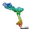
 Citation Citation | Journal: EMBO J / Year: 2011 Title: 4.4 Å cryo-EM structure of an enveloped alphavirus Venezuelan equine encephalitis virus. Authors: Rui Zhang / Corey F Hryc / Yao Cong / Xiangan Liu / Joanita Jakana / Rodion Gorchakov / Matthew L Baker / Scott C Weaver / Wah Chiu /  Abstract: Venezuelan equine encephalitis virus (VEEV), a member of the membrane-containing Alphavirus genus, is a human and equine pathogen, and has been developed as a biological weapon. Using electron cryo- ...Venezuelan equine encephalitis virus (VEEV), a member of the membrane-containing Alphavirus genus, is a human and equine pathogen, and has been developed as a biological weapon. Using electron cryo-microscopy (cryo-EM), we determined the structure of an attenuated vaccine strain, TC-83, of VEEV to 4.4 Å resolution. Our density map clearly resolves regions (including E1, E2 transmembrane helices and cytoplasmic tails) that were missing in the crystal structures of domains of alphavirus subunits. These new features are implicated in the fusion, assembly and budding processes of alphaviruses. Furthermore, our map reveals the unexpected E3 protein, which is cleaved and generally thought to be absent in the mature VEEV. Our structural results suggest a mechanism for the initial stage of nucleocapsid core formation, and shed light on the virulence attenuation, host recognition and neutralizing activities of VEEV and other alphavirus pathogens. | ||||||
| History |
|
- Structure visualization
| Movie |
Movie viewer |
|---|---|
| Structure viewer | Molecule: MolmilJmol/JSmol |
- Downloads & links
-Download
| PDBx/mmCIF format | 3j0g.cif.gz | 48.2 KB | Display | PDBx/mmCIF format |
|---|---|---|---|---|
| PDB format | pdb3j0g.ent.gz | 37.9 KB | Display | PDB format |
| PDBx/mmJSON format | 3j0g.json.gz | Tree view | PDBx/mmJSON format | |
| Others |  Other downloads Other downloads |
-Validation report
| Summary document | 3j0g_validation.pdf.gz | 1.2 MB | Display | wwPDB validaton report |
|---|---|---|---|---|
| Full document | 3j0g_full_validation.pdf.gz | 1.2 MB | Display | |
| Data in XML | 3j0g_validation.xml.gz | 14.2 KB | Display | |
| Data in CIF | 3j0g_validation.cif.gz | 19.6 KB | Display | |
| Arichive directory | https://data.pdbj.org/pub/pdb/validation_reports/j0/3j0gftp://data.pdbj.org/pub/pdb/validation_reports/j0/3j0g | HTTPS FTP |
-Related structure data
-Links
 PDBj
PDBj


- Assembly
| Deposited unit | 
|
|---|---|
| 1 | x 60
|
| 2 |
|
| 3 | x 5
|
| 4 | x 6
|
| 5 | 
|
| Symmetry | Point symmetry: (Schoenflies symbol: I (icosahedral)) |
-Components
| #1: Protein | Mass: 6488.601 Da / Num. of mol.: 4 / Fragment: UNP residues 276-334 / Source method: isolated from a natural source Details: purified from infected Baby hamster kidney (BHK) cells Source: (natural) Venezuelan equine encephalitis virus / Strain: TC-83 / References: UniProt: P05674 |
|---|
-Experimental details
-Experiment
| Experiment | Method: ELECTRON MICROSCOPY |
|---|---|
| EM experiment | Aggregation state: PARTICLE / 3D reconstruction method: single particle reconstruction |
- Sample preparation
| Component | Name: Venezuelan Equine Encephalitis Virus TC83 strain E3 protein that is cleaved from p62 protein Type: VIRUS Details: The virus contains 240 copies of E1, E2, E3 and capsid proteins |
|---|---|
| Details of virus | Empty: NO / Enveloped: YES / Host category: VERTEBRATES / Isolate: STRAIN / Type: VIRION |
| Natural host | Organism: Cricetinae / Strain: Baby Hamster Kidney cells |
| Buffer solution | Name: TEN buffer / pH: 7.4 / Details: TEN buffer |
| Specimen | Embedding applied: NO / Shadowing applied: NO / Staining applied: NO / Vitrification applied: YES |
| Specimen support | Details: two of the eight grids used for imaging contained continuous carbon film underneath the samples |
| Vitrification | Instrument: FEI VITROBOT MARK I / Cryogen name: ETHANE / Temp: 89.34 K / Humidity: 100 % / Details: vitrification carried out in Chiu lab, Houston, TX Method: 2 blots, each blot 2 second before plunging, offset = 0 |
- Electron microscopy imaging
| Microscopy | Model: JEOL 3200FSC / Date: Apr 9, 2008 / Details: omega energy filter |
|---|---|
| Electron gun | Electron source:  FIELD EMISSION GUN / Accelerating voltage: 300 kV / Illumination mode: FLOOD BEAM FIELD EMISSION GUN / Accelerating voltage: 300 kV / Illumination mode: FLOOD BEAM |
| Electron lens | Mode: BRIGHT FIELD / Nominal magnification: 100000 X / Nominal defocus max: 2500 nm / Nominal defocus min: 400 nm / Cs: 4.3 mm Astigmatism: objective lens astigmatism was corrected at 100,000 times magnification |
| Specimen holder | Specimen holder model: JEOL 3200FSC CRYOHOLDER / Specimen holder type: Eucentric / Temperature: 96 K / Tilt angle max: 0 ° / Tilt angle min: 0 ° |
| Image recording | Electron dose: 18 e/Å2 / Film or detector model: GATAN ULTRASCAN 4000 (4k x 4k) |
| EM imaging optics | Energyfilter name: In-column Omega Filter |
- Processing
| EM software |
| ||||||||||||
|---|---|---|---|---|---|---|---|---|---|---|---|---|---|
| CTF correction | Details: CTF parameters were determined from particles within each CCD image | ||||||||||||
| Symmetry | Point symmetry: I (icosahedral) | ||||||||||||
| 3D reconstruction | Method: projection matching / Resolution: 4.8 Å / Num. of particles: 37000 / Nominal pixel size: 1.07 Å Details: After each iteration, the non-icosahedral parts (which included the lipids and the RNA) in the reconstruction were removed by a soft-edged mask derived from an icosahedrally organized, ...Details: After each iteration, the non-icosahedral parts (which included the lipids and the RNA) in the reconstruction were removed by a soft-edged mask derived from an icosahedrally organized, protein-only map that was low-pass filtered to 30 Angstroms. This map then served as the reference model for the next iteration. Num. of class averages: 3328 / Symmetry type: POINT | ||||||||||||
| Atomic model building | Protocol: RIGID BODY FIT / Space: REAL Details: METHOD--rigid-body fitting DETAILS--The E3 homology model was not further refined against the cryo-EM density map due to its less-resolved quality | ||||||||||||
| Atomic model building | PDB-ID: 3N40 | ||||||||||||
| Refinement step | Cycle: LAST
|