 Movie
Movie Controller

[English] 日本語
 Yorodumi
- PDB-3iyx: Coordinates of the b1b bridge-forming protein structures fitted i... -
+ Open data

- Basic information
| Entry | Database: PDB / ID: 3iyx | ||||||
|---|---|---|---|---|---|---|---|
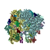
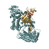
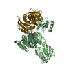
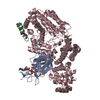
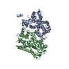
| Title | Coordinates of the b1b bridge-forming protein structures fitted into the Cryo-EM map of E.coli 70S ribosome (EMD-1056) | ||||||
 Components Components |
| ||||||
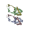
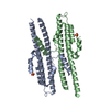
 Keywords Keywords | RIBOSOMAL PROTEIN / Ribosomal intersubunit bridges / B1b-bridge / Ratchet-like motion / Ribosomal protein L31 | ||||||
| Function / homology |  Function and homology information Function and homology informationtRNA binding / rRNA binding / ribosome / structural constituent of ribosome / translation / metal ion binding / cytoplasm Similarity search - Function | ||||||
| Biological species |  | ||||||
| Method | ELECTRON MICROSCOPY / single particle reconstruction / cryo EM / Resolution: 9 Å | ||||||
 Authors Authors | Shasmal, M. / Chakraborty, B. / Sengupta, J. | ||||||
 Citation Citation | Journal: Biochem Biophys Res Commun / Year: 2010 Title: Intrinsic molecular properties of the protein-protein bridge facilitate ratchet-like motion of the ribosome. Authors: Manidip Shasmal / Biprashekhar Chakraborty / Jayati Sengupta /  Abstract: The ribosomal intersubunit bridges maintain the overall architecture of the ribosome and thereby play a pivotal role in the dynamics of translation. The only protein-protein bridge, b1b, is formed by ...The ribosomal intersubunit bridges maintain the overall architecture of the ribosome and thereby play a pivotal role in the dynamics of translation. The only protein-protein bridge, b1b, is formed by the two proteins, S13 and L5 of the small and large ribosomal subunits, respectively. B1b absorbs the largest movement during ratchet-like motion, and its two proteins reorganize in different constellations during this motion of the ribosome. Our results in this study of b1b in the Escherichia coli 70S ribosome suggest that the intrinsic molecular features of the bridging proteins allow the bridge to modulate the ratchet-like motion in a controlled manner. Additionally, another large subunit protein, L31, seems to participate with S13 and L5 in the formation, dynamics, and stabilization of this bridge. | ||||||
| History |
|
- Structure visualization
| Movie |
Movie viewer |
|---|---|
| Structure viewer | Molecule: MolmilJmol/JSmol |
- Downloads & links
-Download
| PDBx/mmCIF format | 3iyx.cif.gz | 24.2 KB | Display | PDBx/mmCIF format |
|---|---|---|---|---|
| PDB format | pdb3iyx.ent.gz | 11.2 KB | Display | PDB format |
| PDBx/mmJSON format | 3iyx.json.gz | Tree view | PDBx/mmJSON format | |
| Others |  Other downloads Other downloads |
-Validation report
| Summary document | 3iyx_validation.pdf.gz | 762.3 KB | Display | wwPDB validaton report |
|---|---|---|---|---|
| Full document | 3iyx_full_validation.pdf.gz | 761.8 KB | Display | |
| Data in XML | 3iyx_validation.xml.gz | 11.5 KB | Display | |
| Data in CIF | 3iyx_validation.cif.gz | 15.5 KB | Display | |
| Arichive directory | https://data.pdbj.org/pub/pdb/validation_reports/iy/3iyxftp://data.pdbj.org/pub/pdb/validation_reports/iy/3iyx | HTTPS FTP |
-Related structure data
| Related structure data | 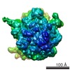 1056M  3iyyC M: map data used to model this data C: citing same article ( |
|---|---|
| Similar structure data |


-Links
 PDBj
PDBj


- Assembly
| Deposited unit | 
|
|---|---|
| 1 |
|
-Components
| #1: Protein | Mass: 13128.467 Da / Num. of mol.: 1 / Source method: isolated from a natural source / Source: (natural) |
|---|---|
| #2: Protein | Mass: 20333.611 Da / Num. of mol.: 1 / Source method: isolated from a natural source / Source: (natural) |
| #3: Protein | Mass: 7887.117 Da / Num. of mol.: 1 / Source method: isolated from a natural source / Source: (natural) |
-Experimental details
-Experiment
| Experiment | Method: ELECTRON MICROSCOPY |
|---|---|
| EM experiment | Aggregation state: PARTICLE / 3D reconstruction method: single particle reconstruction |
- Sample preparation
| Component | Name: E.coli 70S ribosome / Type: RIBOSOME |
|---|---|
| Buffer solution | Name: Polymix buffer / pH: 7.5 / Details: Polymix buffer |
| Specimen | Embedding applied: NO / Shadowing applied: NO / Staining applied: NO / Vitrification applied: YES |
| Specimen support | Details: Quantifoil holley carbon film grids |
| Vitrification | Cryogen name: ETHANE / Details: Rapid-freezing in liquid ethane |
- Electron microscopy imaging
| Experimental equipment |  Model: Tecnai F20 / Image courtesy: FEI Company |
|---|---|
| Microscopy | Model: FEI TECNAI F20 |
| Electron gun | Electron source:  FIELD EMISSION GUN / Accelerating voltage: 200 kV / Illumination mode: FLOOD BEAM FIELD EMISSION GUN / Accelerating voltage: 200 kV / Illumination mode: FLOOD BEAM |
| Electron lens | Mode: BRIGHT FIELD / Nominal magnification: 50000 X / Calibrated magnification: 49696 X / Nominal defocus max: 4000 nm / Nominal defocus min: 2000 nm / Cs: 2 mm |
| Specimen holder | Temperature: 93 K / Tilt angle max: 0 ° / Tilt angle min: 0 ° |
| Image recording | Electron dose: 20 e/Å2 / Film or detector model: KODAK SO-163 FILM |
| Radiation | Protocol: SINGLE WAVELENGTH / Monochromatic (M) / Laue (L): M / Scattering type: x-ray |
| Radiation wavelength | Relative weight: 1 |
- Processing
| EM software | Name: SPIDER / Category: 3D reconstruction | ||||||||||||||||||||||||||||
|---|---|---|---|---|---|---|---|---|---|---|---|---|---|---|---|---|---|---|---|---|---|---|---|---|---|---|---|---|---|
| CTF correction | Details: CTF correction of 3D maps by Wiener filteration | ||||||||||||||||||||||||||||
| Symmetry | Point symmetry: C1 (asymmetric) | ||||||||||||||||||||||||||||
| 3D reconstruction | Method: Single Particle / Resolution: 9 Å / Num. of particles: 52181 / Nominal pixel size: 2.82 Å / Magnification calibration: TMV / Symmetry type: POINT | ||||||||||||||||||||||||||||
| Atomic model building |
| ||||||||||||||||||||||||||||
| Atomic model building | Source name: PDB / Type: experimental model
| ||||||||||||||||||||||||||||
| Refinement step | Cycle: LAST
|

