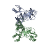
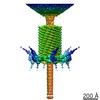
Journal: Structure / Year: 2009 Title: The structure of gene product 6 of bacteriophage T4, the hinge-pin of the baseplate. Authors: Anastasia A Aksyuk / Petr G Leiman / Mikhail M Shneider / Vadim V Mesyanzhinov / Michael G Rossmann / Abstract: The baseplate of bacteriophage T4 is a multicomponent protein complex, which controls phage attachment to the host. It assembles from six wedges and a central hub. During infection the baseplate ...The baseplate of bacteriophage T4 is a multicomponent protein complex, which controls phage attachment to the host. It assembles from six wedges and a central hub. During infection the baseplate undergoes a large conformational change from a dome-shaped to a flat, star-shaped structure. We report the crystal structure of the C-terminal half of gene product (gp) 6 and investigate its motion with respect to the other proteins during the baseplate rearrangement. Six gp6 dimers interdigitate, forming a ring that maintains the integrity of the baseplate in both conformations. One baseplate wedge contains an N-terminal dimer of gp6, whereas neighboring wedges are tied together through the C-terminal dimer of gp6. The dimeric interactions are preserved throughout the rearrangement of the baseplate. However, the hinge angle between the N- and C-terminal parts of gp6 changes by approximately 15 degrees , accounting for a 10 A radial increase in the diameter of the gp6 ring.
History
Deposition
Apr 17, 2009
Deposition site: RCSB / Processing site: RCSB
Revision 1.0
May 19, 2009
Provider: repository / Type: Initial release
Revision 1.1
Jul 13, 2011
Group: Version format compliance
Revision 1.2
Jul 18, 2018
Group: Data collection / Category: em_software / Item: _em_software.image_processing_id
A: Baseplate structural protein Gp6 B: Baseplate structural protein Gp6 C: Baseplate structural protein Gp6 D: Baseplate structural protein Gp6 E: Baseplate structural protein Gp6 F: Baseplate structural protein Gp6 G: Baseplate structural protein Gp6 H: Baseplate structural protein Gp6 I: Baseplate structural protein Gp6 J: Baseplate structural protein Gp6 K: Baseplate structural protein Gp6 L: Baseplate structural protein Gp6
In the structure databanks used in Yorodumi, some data are registered as the other names, "COVID-19 virus" and "2019-nCoV". Here are the details of the virus and the list of structure data.
Jan 31, 2019. EMDB accession codes are about to change! (news from PDBe EMDB page)
EMDB accession codes are about to change! (news from PDBe EMDB page)
The allocation of 4 digits for EMDB accession codes will soon come to an end. Whilst these codes will remain in use, new EMDB accession codes will include an additional digit and will expand incrementally as the available range of codes is exhausted. The current 4-digit format prefixed with “EMD-” (i.e. EMD-XXXX) will advance to a 5-digit format (i.e. EMD-XXXXX), and so on. It is currently estimated that the 4-digit codes will be depleted around Spring 2019, at which point the 5-digit format will come into force.
The EM Navigator/Yorodumi systems omit the EMD- prefix.
Related info.:Q: What is EMD? / ID/Accession-code notation in Yorodumi/EM Navigator
Yorodumi is a browser for structure data from EMDB, PDB, SASBDB, etc.
This page is also the successor to EM Navigator detail page, and also detail information page/front-end page for Omokage search.
The word "yorodu" (or yorozu) is an old Japanese word meaning "ten thousand". "mi" (miru) is to see.
Related info.:EMDB / PDB / SASBDB / Comparison of 3 databanks / Yorodumi Search / Aug 31, 2016. New EM Navigator & Yorodumi / Yorodumi Papers / Jmol/JSmol / Function and homology information / Changes in new EM Navigator and Yorodumi
 Movie
Movie Controller

 Yorodumi
 Open data
 Basic information
 Components
 Keywords
 VIRAL PROTEIN /
  Function and homology information
 Authors
 Citation
 Structure visualization
 Downloads & links
 Other downloads

 PDBj
PDBj Assembly

 Sample preparation
 Electron microscopy imaging
 Processing