[English] 日本語
Yorodumi
- PDB-3h2t: Crystal structure of gene product 6, baseplate protein of bacteri... -

+
Open data


ID or keywords:

Loading...

-
Basic information

Entry
Database: PDB / ID: 3h2t
TitleCrystal structure of gene product 6, baseplate protein of bacteriophage T4
ComponentsBaseplate structural protein Gp6
KeywordsVIRAL PROTEIN / Virion
Function / homology
Function and homology information


virus tail, baseplate / viral tail assembly / identical protein binding
Similarity search - Function
GMP Synthetase; Chain A, domain 3 - #200 / Baseplate wedge protein gp6 / : / : / : / : / Baseplate wedge protein gp6-like, helical domain / Baseplate structural protein gp6, C-terminal domain I / Baseplate structural protein gp6, C-terminal domain II / Baseplate wedge protein gp6, domain II ...GMP Synthetase; Chain A, domain 3 - #200 / Baseplate wedge protein gp6 / : / : / : / : / Baseplate wedge protein gp6-like, helical domain / Baseplate structural protein gp6, C-terminal domain I / Baseplate structural protein gp6, C-terminal domain II / Baseplate wedge protein gp6, domain II / GMP Synthetase; Chain A, domain 3 / 2-Layer Sandwich / Alpha Beta
Similarity search - Domain/homology
Baseplate wedge protein gp6
Similarity search - Component
Biological speciesEnterobacteria phage T4 (virus)
MethodX-RAY DIFFRACTION / SYNCHROTRON / MAD / Resolution: 3.2 Å
AuthorsAksyuk, A.A. / Leiman, P.G. / Shneider, M.M. / Mesyanzhinov, V.V. / Rossmann, M.G.
Citation
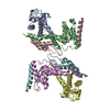
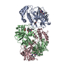
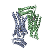
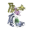
Journal: Structure / Year: 2009
Title: The structure of gene product 6 of bacteriophage T4, the hinge-pin of the baseplate.
Authors: Anastasia A Aksyuk / Petr G Leiman / Mikhail M Shneider / Vadim V Mesyanzhinov / Michael G Rossmann /
Abstract: The baseplate of bacteriophage T4 is a multicomponent protein complex, which controls phage attachment to the host. It assembles from six wedges and a central hub. During infection the baseplate ...The baseplate of bacteriophage T4 is a multicomponent protein complex, which controls phage attachment to the host. It assembles from six wedges and a central hub. During infection the baseplate undergoes a large conformational change from a dome-shaped to a flat, star-shaped structure. We report the crystal structure of the C-terminal half of gene product (gp) 6 and investigate its motion with respect to the other proteins during the baseplate rearrangement. Six gp6 dimers interdigitate, forming a ring that maintains the integrity of the baseplate in both conformations. One baseplate wedge contains an N-terminal dimer of gp6, whereas neighboring wedges are tied together through the C-terminal dimer of gp6. The dimeric interactions are preserved throughout the rearrangement of the baseplate. However, the hinge angle between the N- and C-terminal parts of gp6 changes by approximately 15 degrees , accounting for a 10 A radial increase in the diameter of the gp6 ring.
History
DepositionApr 14, 2009Deposition site: RCSB / Processing site: RCSB
Revision 1.0May 19, 2009Provider: repository / Type: Initial release
Revision 1.1Jul 13, 2011Group: Version format compliance
Revision 1.2Feb 20, 2013Group: Other
Revision 1.3Nov 1, 2017Group: Refinement description / Category: software / Item: _software.classification / _software.name
Revision 1.4Feb 21, 2024Group: Data collection / Database references
Category: chem_comp_atom / chem_comp_bond ...chem_comp_atom / chem_comp_bond / database_2 / struct_ref_seq_dif
Item: _database_2.pdbx_DOI / _database_2.pdbx_database_accession / _struct_ref_seq_dif.details

-
Structure visualization

Structure viewerMolecule:
MolmilJmol/JSmol

Downloads & links

-
Assembly

Deposited unit
A: Baseplate structural protein Gp6
B: Baseplate structural protein Gp6


Theoretical massNumber of molelcules
Total (without water)76,6092
Polymers76,6092
Non-polymers00
Water34219
1


  • Idetical with deposited unit
  • defined by author&software
TypeNameSymmetry operationNumber
identity operation1_555x,y,z1
Buried area2120 Å2
ΔGint-9.9 kcal/mol
Surface area32140 Å2
MethodPISA
Unit cell
Length a, b, c (Å)76.774, 94.579, 136.221
Angle α, β, γ (deg.)90.00, 90.00, 90.00
Int Tables number19
Space group name H-MP212121
DetailsSIX DIMERS FORM A RING IN THE BACTERIOPHAGE T4 BASEPLATE

-
Components

#1: Protein Baseplate structural protein Gp6 / Baseplate wedge protein 6


Mass: 38304.449 Da / Num. of mol.: 2 / Fragment: UNP residues 334-660
Source method: isolated from a genetically manipulated source
Source: (gene. exp.) Enterobacteria phage T4 (virus) / Strain: D / Gene: 6, gene 6 / Plasmid: pET22 / Production host: Escherichia coli (E. coli) / Strain (production host): BL21(DE3) / References: UniProt: P19060
#2: Water ChemComp-HOH / water / Water


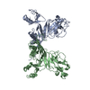
Mass: 18.015 Da / Num. of mol.: 19 / Source method: isolated from a natural source / Formula: H2O

-
Experimental details

-
Experiment

ExperimentMethod: X-RAY DIFFRACTION / Number of used crystals: 2

-
Sample preparation

CrystalDensity Matthews: 3.23 Å3/Da / Density % sol: 61.89 %
Crystal growTemperature: 298 K / Method: vapor diffusion / pH: 7
Details: 1.2 M Ammonium Sulfate, buffered with HEPES, pH 7.0, VAPOR DIFFUSION, temperature 298K

-
Data collection

Diffraction
IDMean temperature (K)Crystal-ID
11001
21001
Diffraction source
SourceSiteBeamlineIDWavelength (Å)
SYNCHROTRONAPS 23-ID-D10.97948
SYNCHROTRONAPS 23-ID-D20.97948
Detector
TypeIDDetectorDate
MARMOSAIC 300 mm CCD1CCDJun 2, 2008
MARMOSAIC 300 mm CCD2CCDFeb 14, 2007
Radiation
IDMonochromatorProtocolMonochromatic (M) / Laue (L)Scattering typeWavelength-ID
1Double crystal cryo-cooled Si(111)SADMx-ray1
2Double crystal cryo-cooled Si(111)SINGLE WAVELENGTHMx-ray1
Radiation wavelengthWavelength: 0.97948 Å / Relative weight: 1
ReflectionResolution: 3.2→50 Å / Num. all: 16941 / Num. obs: 16878 / % possible obs: 99.63 % / Observed criterion σ(I): 2 / Redundancy: 7.2 % / Biso Wilson estimate: 80.95 Å2 / Rmerge(I) obs: 0.096 / Net I/σ(I): 26.9
Reflection shellResolution: 3.2→3.31 Å / Redundancy: 6.3 % / Rmerge(I) obs: 0.476 / Mean I/σ(I) obs: 3.9 / % possible all: 97

-
Processing

Software
NameClassification
HKL-2000data collection
SHELXCDphasing
SHELXEmodel building
PHASERphasing
PHENIXrefinement
HKL-2000data reduction
HKL-2000data scaling
RefinementMethod to determine structure: MAD / Resolution: 3.2→44.856 Å / SU ML: 0.82 / Cross valid method: THROUGHOUT / Stereochemistry target values: MLHL
RfactorNum. reflection% reflectionSelection details
Rfree0.3216 863 5.11 %random
Rwork0.2297 ---
obs0.2344 16878 99.63 %-
Solvent computationShrinkage radii: 0.9 Å / VDW probe radii: 1.11 Å / Solvent model: FLAT BULK SOLVENT MODEL / Bsol: 74.61 Å2 / ksol: 0.337 e/Å3
Displacement parameters
Baniso -1Baniso -2Baniso -3
1-22.7658 Å20 Å20 Å2
2---9.6198 Å2-0 Å2
3----13.146 Å2
Refinement stepCycle: LAST / Resolution: 3.2→44.856 Å
ProteinNucleic acidLigandSolventTotal
Num. atoms5230 0 0 19 5249
Refine LS restraints
Refine-IDTypeDev idealNumber
X-RAY DIFFRACTIONf_bond_d0.0075334
X-RAY DIFFRACTIONf_bond_d_na
X-RAY DIFFRACTIONf_bond_d_prot
X-RAY DIFFRACTIONf_angle_d1.2157242
X-RAY DIFFRACTIONf_angle_d_na
X-RAY DIFFRACTIONf_angle_d_prot
X-RAY DIFFRACTIONf_angle_deg
X-RAY DIFFRACTIONf_angle_deg_na
X-RAY DIFFRACTIONf_angle_deg_prot
X-RAY DIFFRACTIONf_dihedral_angle_d19.1461927
X-RAY DIFFRACTIONf_dihedral_angle_d_na
X-RAY DIFFRACTIONf_dihedral_angle_d_prot
X-RAY DIFFRACTIONf_improper_angle_d0.087813
X-RAY DIFFRACTIONf_improper_angle_d_na
X-RAY DIFFRACTIONf_improper_angle_d_prot
X-RAY DIFFRACTIONf_mcbond_it
X-RAY DIFFRACTIONf_mcangle_it
X-RAY DIFFRACTIONf_scbond_it
X-RAY DIFFRACTIONf_scangle_it
LS refinement shell

Refine-ID: X-RAY DIFFRACTION / Total num. of bins used: 6

Resolution (Å)Rfactor RfreeNum. reflection RfreeRfactor RworkNum. reflection Rwork% reflection obs (%)
3.2-3.40040.39421330.2705260199
3.4004-3.66290.36341450.23942621100
3.6629-4.03130.31611530.22022623100
4.0313-4.61410.26321430.1922652100
4.6141-5.81130.28211430.19972698100
5.8113-44.860.30941460.22372820100

+
About Yorodumi

-
News

-
Feb 9, 2022. New format data for meta-information of EMDB entries

New format data for meta-information of EMDB entries

  • Version 3 of the EMDB header file is now the official format.
  • The previous official version 1.9 will be removed from the archive.

Related info.:EMDB header

External links:wwPDB to switch to version 3 of the EMDB data model

-
Aug 12, 2020. Covid-19 info

Covid-19 info

URL: https://pdbj.org/emnavi/covid19.php

New page: Covid-19 featured information page in EM Navigator.

Related info.:Covid-19 info / Mar 5, 2020. Novel coronavirus structure data

+
Mar 5, 2020. Novel coronavirus structure data

Novel coronavirus structure data

Related info.:Yorodumi Speices / Aug 12, 2020. Covid-19 info

External links:COVID-19 featured content - PDBj / Molecule of the Month (242):Coronavirus Proteases

+
Jan 31, 2019. EMDB accession codes are about to change! (news from PDBe EMDB page)

EMDB accession codes are about to change! (news from PDBe EMDB page)

  • The allocation of 4 digits for EMDB accession codes will soon come to an end. Whilst these codes will remain in use, new EMDB accession codes will include an additional digit and will expand incrementally as the available range of codes is exhausted. The current 4-digit format prefixed with “EMD-” (i.e. EMD-XXXX) will advance to a 5-digit format (i.e. EMD-XXXXX), and so on. It is currently estimated that the 4-digit codes will be depleted around Spring 2019, at which point the 5-digit format will come into force.
  • The EM Navigator/Yorodumi systems omit the EMD- prefix.

Related info.:Q: What is EMD? / ID/Accession-code notation in Yorodumi/EM Navigator

External links:EMDB Accession Codes are Changing Soon! / Contact to PDBj

+
Jul 12, 2017. Major update of PDB

Major update of PDB

  • wwPDB released updated PDB data conforming to the new PDBx/mmCIF dictionary.
  • This is a major update changing the version number from 4 to 5, and with Remediation, in which all the entries are updated.
  • In this update, many items about electron microscopy experimental information are reorganized (e.g. em_software).
  • Now, EM Navigator and Yorodumi are based on the updated data.

External links:wwPDB Remediation / Enriched Model Files Conforming to OneDep Data Standards Now Available in the PDB FTP Archive

-
Yorodumi

Thousand views of thousand structures

  • Yorodumi is a browser for structure data from EMDB, PDB, SASBDB, etc.
  • This page is also the successor to EM Navigator detail page, and also detail information page/front-end page for Omokage search.
  • The word "yorodu" (or yorozu) is an old Japanese word meaning "ten thousand". "mi" (miru) is to see.

Related info.:EMDB / PDB / SASBDB / Comparison of 3 databanks / Yorodumi Search / Aug 31, 2016. New EM Navigator & Yorodumi / Yorodumi Papers / Jmol/JSmol / Function and homology information / Changes in new EM Navigator and Yorodumi

Read more