 Movie
Movie Controller


+ Open data

- Basic information
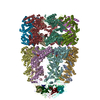
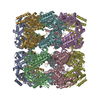
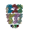
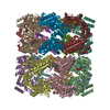
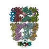
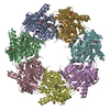
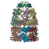
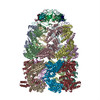
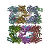
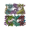
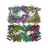
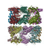
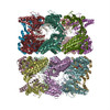
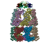
| Entry | Database: PDB / ID: 2cgt | ||||||
|---|---|---|---|---|---|---|---|
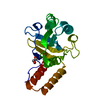
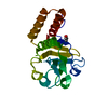
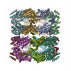
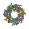
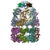
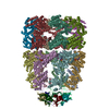
| Title | GROEL-ADP-gp31 COMPLEX | ||||||
 Components Components |
| ||||||
 Keywords Keywords | CHAPERONE / CHAPERONIN / CELL CYCLE / CELL DIVISION / CAPSID ASSEMBLY / EARLY PROTEIN | ||||||
| Function / homology |  Function and homology information Function and homology informationviral capsid assembly / GroEL-GroES complex / chaperonin ATPase / virion assembly / chaperone cofactor-dependent protein refolding / protein folding chaperone / isomerase activity / ATP-dependent protein folding chaperone / response to radiation / unfolded protein binding ...viral capsid assembly / GroEL-GroES complex / chaperonin ATPase / virion assembly / chaperone cofactor-dependent protein refolding / protein folding chaperone / isomerase activity / ATP-dependent protein folding chaperone / response to radiation / unfolded protein binding / protein folding / protein refolding / response to heat / magnesium ion binding / ATP hydrolysis activity / ATP binding / identical protein binding / membrane / cytosol Similarity search - Function | ||||||
| Biological species |   BACTERIOPHAGE T4 (virus) BACTERIOPHAGE T4 (virus) | ||||||
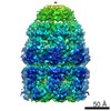
| Method | ELECTRON MICROSCOPY / single particle reconstruction / cryo EM / Resolution: 8.2 Å | ||||||
 Authors Authors | Clare, D.K. / Bakkes, P.J. / van Heerikhuizen, H. / van der Vies, S.M. / Saibil, H.R. | ||||||
 Citation Citation | Journal: J Mol Biol / Year: 2006 Title: An expanded protein folding cage in the GroEL-gp31 complex. Authors: Daniel K Clare / Patrick J Bakkes / Harm van Heerikhuizen / Saskia M van der Vies / Helen R Saibil /  Abstract: Bacteriophage T4 produces a GroES analogue, gp31, which cooperates with the Escherichia coli GroEL to fold its major coat protein gp23. We have used cryo-electron microscopy and image processing to ...Bacteriophage T4 produces a GroES analogue, gp31, which cooperates with the Escherichia coli GroEL to fold its major coat protein gp23. We have used cryo-electron microscopy and image processing to obtain three-dimensional structures of the E.coli chaperonin GroEL complexed with gp31, in the presence of both ATP and ADP. The GroEL-gp31-ADP map has a resolution of 8.2 A, which allows accurate fitting of the GroEL and gp31 crystal structures. Comparison of this fitted structure with that of the GroEL-GroES-ADP structure previously determined by cryo-electron microscopy shows that the folding cage is expanded. The enlarged volume for folding is consistent with the size of the bacteriophage coat protein gp23, which is the major substrate of GroEL-gp31 chaperonin complex. At 56 kDa, gp23 is close to the maximum size limit of a polypeptide that is thought to fit inside the GroEL-GroES folding cage. | ||||||
| History |
| ||||||
| Remark 700 | SHEET THE SHEET STRUCTURE OF THIS MOLECULE IS BIFURCATED. IN ORDER TO REPRESENT THIS FEATURE IN ... SHEET THE SHEET STRUCTURE OF THIS MOLECULE IS BIFURCATED. IN ORDER TO REPRESENT THIS FEATURE IN THE SHEET RECORDS BELOW, TWO SHEETS ARE DEFINED. |
- Structure visualization
| Movie |
Movie viewer |
|---|---|
| Structure viewer | Molecule: MolmilJmol/JSmol |
- Downloads & links
-Download
| PDBx/mmCIF format | 2cgt.cif.gz | 1.3 MB | Display | PDBx/mmCIF format |
|---|---|---|---|---|
| PDB format | pdb2cgt.ent.gz | 1.1 MB | Display | PDB format |
| PDBx/mmJSON format | 2cgt.json.gz | Tree view | PDBx/mmJSON format | |
| Others |  Other downloads Other downloads |
-Validation report
| Summary document | 2cgt_validation.pdf.gz | 1 MB | Display | wwPDB validaton report |
|---|---|---|---|---|
| Full document | 2cgt_full_validation.pdf.gz | 1.3 MB | Display | |
| Data in XML | 2cgt_validation.xml.gz | 223.6 KB | Display | |
| Data in CIF | 2cgt_validation.cif.gz | 328.9 KB | Display | |
| Arichive directory | https://data.pdbj.org/pub/pdb/validation_reports/cg/2cgtftp://data.pdbj.org/pub/pdb/validation_reports/cg/2cgt | HTTPS FTP |
-Related structure data
| Related structure data | 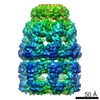 1202MC  1203C M: map data used to model this data C: citing same article ( |
|---|---|
| Similar structure data |


-Links
 PDBj
PDBj

- Assembly
| Deposited unit | 
|
|---|---|
| 1 |
|
-Components
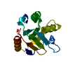
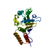
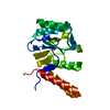
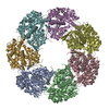
| #1: Protein | Mass: 57260.504 Da / Num. of mol.: 14 Source method: isolated from a genetically manipulated source Details: A TETRADECAMER ARRANGED AS TWO BACK-TO-BACK HEPTAMERS Source: (gene. exp.) #2: Protein | Mass: 12091.999 Da / Num. of mol.: 7 Source method: isolated from a genetically manipulated source Details: HEPTAMER / Source: (gene. exp.) BACTERIOPHAGE T4 (virus) / Plasmid: PAR1 / Production host: Sequence details | RESIDUES 23-46 HAVE BEEN REMOVED FROM THE GP31 STRUCTURE AS THEY WERE IN A DISTORTED CONFORMATION ...RESIDUES 23-46 HAVE BEEN REMOVED FROM THE GP31 STRUCTURE AS THEY WERE IN A DISTORTED CONFORMATI | |
|---|
-Experimental details
-Experiment
| Experiment | Method: ELECTRON MICROSCOPY |
|---|---|
| EM experiment | Aggregation state: PARTICLE / 3D reconstruction method: single particle reconstruction |
- Sample preparation
| Component | Name: GROEL-GP31-ADP / Type: COMPLEX |
|---|---|
| Buffer solution | Name: 20MM TRIS-HCL, 10MM MGCL, 10MM KCL / pH: 7.4 / Details: 20MM TRIS-HCL, 10MM MGCL, 10MM KCL |
| Specimen | Conc.: 1 mg/ml / Embedding applied: NO / Shadowing applied: NO / Staining applied: NO / Vitrification applied: YES |
| Specimen support | Details: HOLEY CARBON |
| Vitrification | Instrument: HOMEMADE PLUNGER / Cryogen name: ETHANE Details: GRIDS WERE BLOTED FOR 2-3 SECONDS AND THEN LEFT TO EQUILIBRATE FOR 2-3 SECONDS AND THEN PLUNGED INTO LIQUID ETHANE |
- Electron microscopy imaging
| Experimental equipment |  Model: Tecnai F20 / Image courtesy: FEI Company |
|---|---|
| Microscopy | Model: FEI TECNAI F20 / Date: Sep 28, 2004 |
| Electron gun | Electron source:  FIELD EMISSION GUN / Accelerating voltage: 200 kV / Illumination mode: FLOOD BEAM FIELD EMISSION GUN / Accelerating voltage: 200 kV / Illumination mode: FLOOD BEAM |
| Electron lens | Mode: BRIGHT FIELD / Nominal magnification: 50000 X / Calibrated magnification: 50000 X / Nominal defocus max: 3300 nm / Nominal defocus min: 1300 nm / Cs: 2 mm |
| Specimen holder | Temperature: 100 K |
| Image recording | Electron dose: 15 e/Å2 / Film or detector model: KODAK SO-163 FILM |
| Image scans | Num. digital images: 28 |
| Radiation wavelength | Relative weight: 1 |
- Processing
| EM software |
| |||||||||||||||||||||
|---|---|---|---|---|---|---|---|---|---|---|---|---|---|---|---|---|---|---|---|---|---|---|
| CTF correction | Details: FULL CORRECTION ON 2D CLASS AVERAGES | |||||||||||||||||||||
| Symmetry | Point symmetry: D7 (2x7 fold dihedral) | |||||||||||||||||||||
| 3D reconstruction | Method: PROJECTION MATCHING / Resolution: 8.2 Å / Num. of particles: 10300 / Nominal pixel size: 1.4 Å / Actual pixel size: 1.4 Å Details: THE 3 DOMAINS OF TWO GROEL SUBUNITS AND A SINGLE GP31 SUBUNIT WERE DOCKED AS RIGID BODIES INTO THE DENSITY MAP Symmetry type: POINT | |||||||||||||||||||||
| Atomic model building | Protocol: RIGID BODY FIT / Space: REAL / Target criteria: Cross-correlation coefficient / Details: METHOD--RIGID BODY REFINEMENT PROTOCOL--X-RAY | |||||||||||||||||||||
| Atomic model building |
| |||||||||||||||||||||
| Refinement | Highest resolution: 8.2 Å | |||||||||||||||||||||
| Refinement step | Cycle: LAST / Highest resolution: 8.2 Å
|

