 Movie
Movie Controller

[English] 日本語
 Yorodumi
- PDB-1aon: CRYSTAL STRUCTURE OF THE ASYMMETRIC CHAPERONIN COMPLEX GROEL/GROE... -
+ Open data

- Basic information
| Entry | Database: PDB / ID: 1aon | ||||||
|---|---|---|---|---|---|---|---|
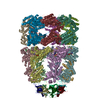
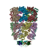
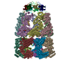
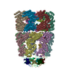
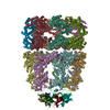
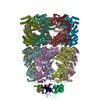
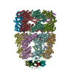
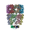
| Title | CRYSTAL STRUCTURE OF THE ASYMMETRIC CHAPERONIN COMPLEX GROEL/GROES/(ADP)7 | ||||||
 Components Components |
| ||||||
 Keywords Keywords | COMPLEX (GROEL/GROES) / COMPLEX (GROEL-GROES) / CHAPERONIN ASSISTED PROTEIN FOLDING / COMPLEX (GROEL-GROES) complex | ||||||
| Function / homology |  Function and homology information Function and homology informationGroEL-GroES complex / chaperonin ATPase / virion assembly / chaperone cofactor-dependent protein refolding / protein folding chaperone / isomerase activity / ATP-dependent protein folding chaperone / response to radiation / unfolded protein binding / protein folding ...GroEL-GroES complex / chaperonin ATPase / virion assembly / chaperone cofactor-dependent protein refolding / protein folding chaperone / isomerase activity / ATP-dependent protein folding chaperone / response to radiation / unfolded protein binding / protein folding / protein-folding chaperone binding / protein refolding / response to heat / magnesium ion binding / ATP hydrolysis activity / ATP binding / identical protein binding / membrane / metal ion binding / cytosol Similarity search - Function | ||||||
| Biological species |  | ||||||
| Method |  X-RAY DIFFRACTION / SYNCHROTRON / MOLECULAR REPLACEMENT / Resolution: 3 Å X-RAY DIFFRACTION / SYNCHROTRON / MOLECULAR REPLACEMENT / Resolution: 3 Å | ||||||
 Authors Authors | Xu, Z. / Horwich, A.L. / Sigler, P.B. | ||||||
 Citation Citation | Journal: Nature / Year: 1997 Title: The crystal structure of the asymmetric GroEL-GroES-(ADP)7 chaperonin complex. Authors: Z Xu / A L Horwich / P B Sigler /  Abstract: Chaperonins assist protein folding with the consumption of ATP. They exist as multi-subunit protein assemblies comprising rings of subunits stacked back to back. In Escherichia coli, asymmetric ...Chaperonins assist protein folding with the consumption of ATP. They exist as multi-subunit protein assemblies comprising rings of subunits stacked back to back. In Escherichia coli, asymmetric intermediates of GroEL are formed with the co-chaperonin GroES and nucleotides bound only to one of the seven-subunit rings (the cis ring) and not to the opposing ring (the trans ring). The structure of the GroEL-GroES-(ADP)7 complex reveals how large en bloc movements of the cis ring's intermediate and apical domains enable bound GroES to stabilize a folding chamber with ADP confined to the cis ring. Elevation and twist of the apical domains double the volume of the central cavity and bury hydrophobic peptide-binding residues in the interface with GroES, as well as between GroEL subunits, leaving a hydrophilic cavity lining that is conducive to protein folding. An inward tilt of the cis equatorial domain causes an outward tilt in the trans ring that opposes the binding of a second GroES. When combined with new functional results, this negative allosteric mechanism suggests a model for an ATP-driven folding cycle that requires a double toroid. #1: Journal: Nature / Year: 1997Title: Distinct Actions of Cis and Trans ATP within the Double Ring of the Chaperonin Groel Authors: Rye, H.S. / Burston, S.G. / Fenton, W.A. / Beechem, J.M. / Xu, Z. / Sigler, P.B. / Horwich, A.L. | ||||||
| History |
|
- Structure visualization
| Structure viewer | Molecule: MolmilJmol/JSmol |
|---|
- Downloads & links
-Download
| PDBx/mmCIF format | 1aon.cif.gz | 1.4 MB | Display | PDBx/mmCIF format |
|---|---|---|---|---|
| PDB format | pdb1aon.ent.gz | 1.1 MB | Display | PDB format |
| PDBx/mmJSON format | 1aon.json.gz | Tree view | PDBx/mmJSON format | |
| Others |  Other downloads Other downloads |
-Validation report
| Summary document | 1aon_validation.pdf.gz | 2.6 MB | Display | wwPDB validaton report |
|---|---|---|---|---|
| Full document | 1aon_full_validation.pdf.gz | 3.2 MB | Display | |
| Data in XML | 1aon_validation.xml.gz | 351.8 KB | Display | |
| Data in CIF | 1aon_validation.cif.gz | 467.4 KB | Display | |
| Arichive directory | https://data.pdbj.org/pub/pdb/validation_reports/ao/1aonftp://data.pdbj.org/pub/pdb/validation_reports/ao/1aon | HTTPS FTP |
-Related structure data
| Related structure data | 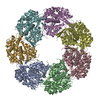 1oelS S: Starting model for refinement |
|---|---|
| Similar structure data |
-Links
 PDBj
PDBj

- Assembly
| Deposited unit | 
| ||||||||||||||||||||||||||||||||||||||||||||||||||||||||||||||||||||||||||||
|---|---|---|---|---|---|---|---|---|---|---|---|---|---|---|---|---|---|---|---|---|---|---|---|---|---|---|---|---|---|---|---|---|---|---|---|---|---|---|---|---|---|---|---|---|---|---|---|---|---|---|---|---|---|---|---|---|---|---|---|---|---|---|---|---|---|---|---|---|---|---|---|---|---|---|---|---|---|
| 1 |
| ||||||||||||||||||||||||||||||||||||||||||||||||||||||||||||||||||||||||||||
| Unit cell |
| ||||||||||||||||||||||||||||||||||||||||||||||||||||||||||||||||||||||||||||
| Noncrystallographic symmetry (NCS) | NCS oper:
|
-Components
| #1: Protein | Mass: 57260.504 Da / Num. of mol.: 14 Source method: isolated from a genetically manipulated source Source: (gene. exp.) #2: Protein | Mass: 10400.938 Da / Num. of mol.: 7 Source method: isolated from a genetically manipulated source Source: (gene. exp.) #3: Chemical | ChemComp-MG /   Mass: 24.305 Da / Num. of mol.: 7 / Source method: obtained synthetically / Formula: Mg Mass: 24.305 Da / Num. of mol.: 7 / Source method: obtained synthetically / Formula: Mg#4: Chemical | ChemComp-ADP /   Mass: 427.201 Da / Num. of mol.: 7 / Source method: obtained synthetically / Formula: C10H15N5O10P2 / Comment: ADP, energy-carrying molecule*YM Mass: 427.201 Da / Num. of mol.: 7 / Source method: obtained synthetically / Formula: C10H15N5O10P2 / Comment: ADP, energy-carrying molecule*YM |
|---|
-Experimental details
-Experiment
| Experiment | Method: X-RAY DIFFRACTION / Number of used crystals: 1 |
|---|
- Sample preparation
| Crystal | Density Matthews: 3.5 Å3/Da / Density % sol: 65 % | ||||||||||||||||||||||||||||||||||||||||
|---|---|---|---|---|---|---|---|---|---|---|---|---|---|---|---|---|---|---|---|---|---|---|---|---|---|---|---|---|---|---|---|---|---|---|---|---|---|---|---|---|---|
| Crystal grow | pH: 5.5 Details: PROTEIN WAS CRYSTALLIZED FROM 12% PEG3000, 0.25M SODIUM GLUTAMATE, 100MM CACODYLIC ACID, PH 5.5 | ||||||||||||||||||||||||||||||||||||||||
| Crystal grow | *PLUS Temperature: 18 ℃ / Method: vapor diffusion, hanging drop / Details: used to seeding | ||||||||||||||||||||||||||||||||||||||||
| Components of the solutions | *PLUS
|
-Data collection
| Diffraction | Mean temperature: 100 K |
|---|---|
| Diffraction source | Source: SYNCHROTRON / Site: NSLS / Beamline: X25 / Wavelength: 0.95 |
| Detector | Type: MARRESEARCH / Detector: IMAGE PLATE / Date: Sep 1, 1996 / Details: MIRRORS |
| Radiation | Monochromator: TWO CRYSTAL NON-DISPERSIVE / Monochromatic (M) / Laue (L): M / Scattering type: x-ray |
| Radiation wavelength | Wavelength: 0.95 Å / Relative weight: 1 |
| Reflection | Resolution: 3→99 Å / Num. obs: 242684 / % possible obs: 96.7 % / Redundancy: 3.3 % / Rsym value: 0.121 / Net I/σ(I): 9.1 |
| Reflection shell | Resolution: 3→3.14 Å / Redundancy: 2.4 % / Mean I/σ(I) obs: 1.8 / Rsym value: 0.53 / % possible all: 91.2 |
| Reflection | *PLUS Rmerge(I) obs: 0.121 |
| Reflection shell | *PLUS % possible obs: 91.2 % / Num. unique obs: 28317 / Rmerge(I) obs: 0.53 |
- Processing
| Software |
| ||||||||||||||||||||||||||||||||||||||||||||||||||||||||||||||||||||||||||||||||
|---|---|---|---|---|---|---|---|---|---|---|---|---|---|---|---|---|---|---|---|---|---|---|---|---|---|---|---|---|---|---|---|---|---|---|---|---|---|---|---|---|---|---|---|---|---|---|---|---|---|---|---|---|---|---|---|---|---|---|---|---|---|---|---|---|---|---|---|---|---|---|---|---|---|---|---|---|---|---|---|---|---|
| Refinement | Method to determine structure: MOLECULAR REPLACEMENT Starting model: PDB ENTRY 1OEL Resolution: 3→40 Å / Data cutoff high absF: 1000000 / Data cutoff low absF: 0.001 / Isotropic thermal model: RESTRAINED / Cross valid method: THROUGHOUT / σ(F): 2 Details: RESIDUE 526 - 547 IN EACH GROEL CHAIN ARE ABSENT FROM THE MODEL BECAUSE THE ELECTRON DENSITY FOR THESE RESIDUES COULD NOT BE INTERPRETED. ARG 197, PHE 204, LYS 225, LYS 226, SER 228 - GLU ...Details: RESIDUE 526 - 547 IN EACH GROEL CHAIN ARE ABSENT FROM THE MODEL BECAUSE THE ELECTRON DENSITY FOR THESE RESIDUES COULD NOT BE INTERPRETED. ARG 197, PHE 204, LYS 225, LYS 226, SER 228 - GLU 232, GLU 238 IN CHAINS A - G ARE MODELLED AS ALANINE.
| ||||||||||||||||||||||||||||||||||||||||||||||||||||||||||||||||||||||||||||||||
| Displacement parameters | Biso mean: 61.5 Å2
| ||||||||||||||||||||||||||||||||||||||||||||||||||||||||||||||||||||||||||||||||
| Refine analyze |
| ||||||||||||||||||||||||||||||||||||||||||||||||||||||||||||||||||||||||||||||||
| Refinement step | Cycle: LAST / Resolution: 3→40 Å
| ||||||||||||||||||||||||||||||||||||||||||||||||||||||||||||||||||||||||||||||||
| Refine LS restraints |
| ||||||||||||||||||||||||||||||||||||||||||||||||||||||||||||||||||||||||||||||||
| Refine LS restraints NCS | NCS model details: RESTRAINTS | ||||||||||||||||||||||||||||||||||||||||||||||||||||||||||||||||||||||||||||||||
| LS refinement shell | Resolution: 3→3.14 Å / Total num. of bins used: 8
| ||||||||||||||||||||||||||||||||||||||||||||||||||||||||||||||||||||||||||||||||
| Xplor file |
| ||||||||||||||||||||||||||||||||||||||||||||||||||||||||||||||||||||||||||||||||
| Software | *PLUS Name: X-PLOR / Version: 3.861 / Classification: refinement | ||||||||||||||||||||||||||||||||||||||||||||||||||||||||||||||||||||||||||||||||
| Refine LS restraints | *PLUS
|
