 Movie
Movie Controller

[English] 日本語
 Yorodumi
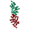
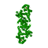
- PDB-1rdw: Actin Crystal Dynamics: Structural Implications for F-actin Nucle... -
+ Open data

- Basic information
| Entry | Database: PDB / ID: 1rdw | ||||||
|---|---|---|---|---|---|---|---|
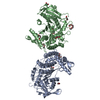
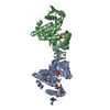
| Title | Actin Crystal Dynamics: Structural Implications for F-actin Nucleation, Polymerization and Branching Mediated by the Anti-parallel Dimer | ||||||
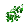
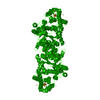
 Components Components | Actin, alpha skeletal muscle | ||||||
 Keywords Keywords | STRUCTURAL PROTEIN / Anti-parallel dimer / Polymerization / Nucleation / Filament | ||||||
| Function / homology |  Function and homology information Function and homology informationcytoskeletal motor activator activity / tropomyosin binding / mesenchyme migration / troponin I binding / myosin heavy chain binding / filamentous actin / actin filament bundle / skeletal muscle thin filament assembly / striated muscle thin filament / actin filament bundle assembly ...cytoskeletal motor activator activity / tropomyosin binding / mesenchyme migration / troponin I binding / myosin heavy chain binding / filamentous actin / actin filament bundle / skeletal muscle thin filament assembly / striated muscle thin filament / actin filament bundle assembly / skeletal muscle myofibril / actin monomer binding / skeletal muscle fiber development / stress fiber / titin binding / actin filament polymerization / filopodium / actin filament / Hydrolases; Acting on acid anhydrides; Acting on acid anhydrides to facilitate cellular and subcellular movement / calcium-dependent protein binding / lamellipodium / cell body / hydrolase activity / protein domain specific binding / calcium ion binding / positive regulation of gene expression / magnesium ion binding / ATP binding / identical protein binding / cytoplasm Similarity search - Function | ||||||
| Biological species |  | ||||||
| Method |  X-RAY DIFFRACTION / MOLECULAR REPLACEMENT / Resolution: 2.3 Å X-RAY DIFFRACTION / MOLECULAR REPLACEMENT / Resolution: 2.3 Å | ||||||
 Authors Authors | Reutzel, R. / Yoshioka, C. / Govindasamy, L. / Yarmola, E.G. / Agbandje-Mckenna, M. / Bubb, M.R. / Mckenna, R. | ||||||
 Citation Citation | Journal: J.Struct.Biol. / Year: 2004 Title: Actin crystal dynamics: structural implications for F-actin nucleation, polymerization, and branching mediated by the anti-parallel dimer. Authors: Reutzel, R. / Yoshioka, C. / Govindasamy, L. / Yarmola, E.G. / Agbandje-McKenna, M. / Bubb, M.R. / McKenna, R. | ||||||
| History |
|
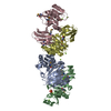
- Structure visualization
| Structure viewer | Molecule: MolmilJmol/JSmol |
|---|
- Downloads & links
-Download
| PDBx/mmCIF format | 1rdw.cif.gz | 90.3 KB | Display | PDBx/mmCIF format |
|---|---|---|---|---|
| PDB format | pdb1rdw.ent.gz | 67.3 KB | Display | PDB format |
| PDBx/mmJSON format | 1rdw.json.gz | Tree view | PDBx/mmJSON format | |
| Others |  Other downloads Other downloads |
-Validation report
| Summary document | 1rdw_validation.pdf.gz | 537.5 KB | Display | wwPDB validaton report |
|---|---|---|---|---|
| Full document | 1rdw_full_validation.pdf.gz | 550.9 KB | Display | |
| Data in XML | 1rdw_validation.xml.gz | 11.4 KB | Display | |
| Data in CIF | 1rdw_validation.cif.gz | 16.7 KB | Display | |
| Arichive directory | https://data.pdbj.org/pub/pdb/validation_reports/rd/1rdwftp://data.pdbj.org/pub/pdb/validation_reports/rd/1rdw | HTTPS FTP |
-Related structure data
| Related structure data | 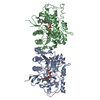 1rfqC 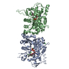 1lcuS S: Starting model for refinement C: citing same article ( |
|---|---|
| Similar structure data |
-Links
 PDBj
PDBj


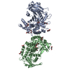
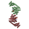
- Assembly
| Deposited unit | 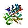
| ||||||||
|---|---|---|---|---|---|---|---|---|---|
| 1 | 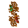
| ||||||||
| Unit cell |
| ||||||||
| Components on special symmetry positions |
|
-Components
| #1: Protein | Mass: 41862.613 Da / Num. of mol.: 1 / Fragment: ACtin Source method: isolated from a genetically manipulated source Source: (gene. exp.) | ||||||
|---|---|---|---|---|---|---|---|
| #2: Chemical |   Mass: 24.305 Da / Num. of mol.: 2 / Source method: obtained synthetically / Formula: Mg Mass: 24.305 Da / Num. of mol.: 2 / Source method: obtained synthetically / Formula: Mg#3: Chemical | ChemComp-ATP / |   Mass: 507.181 Da / Num. of mol.: 1 / Source method: obtained synthetically / Formula: C10H16N5O13P3 / Comment: ATP, energy-carrying molecule*YM Mass: 507.181 Da / Num. of mol.: 1 / Source method: obtained synthetically / Formula: C10H16N5O13P3 / Comment: ATP, energy-carrying molecule*YM#4: Chemical | ChemComp-LAR / | 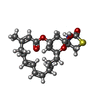  Mass: 421.550 Da / Num. of mol.: 1 / Source method: obtained synthetically / Formula: C22H31NO5S / Comment: toxin*YM Mass: 421.550 Da / Num. of mol.: 1 / Source method: obtained synthetically / Formula: C22H31NO5S / Comment: toxin*YM#5: Water | ChemComp-HOH / |  Mass: 18.015 Da / Num. of mol.: 113 / Source method: isolated from a natural source / Formula: H2O Mass: 18.015 Da / Num. of mol.: 113 / Source method: isolated from a natural source / Formula: H2O |
-Experimental details
-Experiment
| Experiment | Method: X-RAY DIFFRACTION / Number of used crystals: 1 |
|---|
- Sample preparation
| Crystal | Density Matthews: 3.16 Å3/Da / Density % sol: 61.04 % | |||||||||||||||||||||||||||||||||||
|---|---|---|---|---|---|---|---|---|---|---|---|---|---|---|---|---|---|---|---|---|---|---|---|---|---|---|---|---|---|---|---|---|---|---|---|---|
| Crystal grow | Method: vapor diffusion, hanging drop / pH: 6.7 Details: MgCl2, NH4SO4, pH 6.7, VAPOR DIFFUSION, HANGING DROP | |||||||||||||||||||||||||||||||||||
| Crystal grow | *PLUS Temperature: 4 ℃ / Method: vapor diffusion / Details: Bubb, M.R., (2002) J. Biol. Chem., 277, 20999. | |||||||||||||||||||||||||||||||||||
| Components of the solutions | *PLUS
|
-Data collection
| Diffraction | Mean temperature: 273 K |
|---|---|
| Diffraction source | Source: ROTATING ANODE / Type: RIGAKU / Wavelength: 1.5418 Å |
| Detector | Type: RIGAKU RAXIS IV / Detector: IMAGE PLATE / Details: Osmic mirrors |
| Radiation | Monochromator: CuKalpha / Protocol: SINGLE WAVELENGTH / Monochromatic (M) / Laue (L): M / Scattering type: x-ray |
| Radiation wavelength | Wavelength: 1.5418 Å / Relative weight: 1 |
| Reflection | Resolution: 2.3→20 Å / Num. obs: 22951 / % possible obs: 93.9 % / Rsym value: 0.093 |
| Reflection shell | Resolution: 2.3→2.38 Å / Num. unique all: 22951 / Rsym value: 0.424 / % possible all: 92.1 |
| Reflection | *PLUS Rmerge(I) obs: 0.093 |
- Processing
| Software |
| ||||||||||||||||
|---|---|---|---|---|---|---|---|---|---|---|---|---|---|---|---|---|---|
| Refinement | Method to determine structure: MOLECULAR REPLACEMENT Starting model: PDB entry 1LCU Resolution: 2.3→20 Å / Stereochemistry target values: Engh & Huber
| ||||||||||||||||
| Displacement parameters | Biso mean: 38.346 Å2 | ||||||||||||||||
| Refinement step | Cycle: LAST / Resolution: 2.3→20 Å
| ||||||||||||||||
| Refine LS restraints |
| ||||||||||||||||
| Software | *PLUS Version: 5 / Classification: refinement | ||||||||||||||||
| Refinement | *PLUS % reflection Rfree: 5 % | ||||||||||||||||
| Solvent computation | *PLUS | ||||||||||||||||
| Displacement parameters | *PLUS | ||||||||||||||||
| Refine LS restraints | *PLUS
|
