Movie
Movie Controller

[English] 日本語
 Yorodumi
- PDB-1mj1: FITTING THE TERNARY COMPLEX OF EF-Tu/tRNA/GTP AND RIBOSOMAL PROTE... -
+ Open data

- Basic information
| Entry | Database: PDB / ID: 1mj1 | ||||||
|---|---|---|---|---|---|---|---|
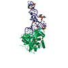
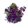
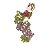
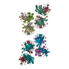
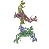
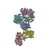
| Title | FITTING THE TERNARY COMPLEX OF EF-Tu/tRNA/GTP AND RIBOSOMAL PROTEINS INTO A 13 A CRYO-EM MAP OF THE COLI 70S RIBOSOME | ||||||
 Components Components |
| ||||||
 Keywords Keywords | RIBOSOME / 70S RIBOSOME / LOW RESOLUTION MODEL TERNARY COMPLEX / EF-Tu | ||||||
| Function / homology |  Function and homology information Function and homology informationendo-alpha-N-acetylgalactosaminidase activity / misfolded RNA binding / Group I intron splicing / RNA folding / Hydrolases; Glycosylases; Glycosidases, i.e. enzymes that hydrolyse O- and S-glycosyl compounds / translation elongation factor activity / positive regulation of RNA splicing / maintenance of translational fidelity / small ribosomal subunit / large ribosomal subunit rRNA binding ...endo-alpha-N-acetylgalactosaminidase activity / misfolded RNA binding / Group I intron splicing / RNA folding / Hydrolases; Glycosylases; Glycosidases, i.e. enzymes that hydrolyse O- and S-glycosyl compounds / translation elongation factor activity / positive regulation of RNA splicing / maintenance of translational fidelity / small ribosomal subunit / large ribosomal subunit rRNA binding / cytosolic small ribosomal subunit / cytosolic large ribosomal subunit / cytoplasmic translation / tRNA binding / rRNA binding / ribosome / structural constituent of ribosome / translation / response to antibiotic / GTPase activity / GTP binding / metal ion binding / cytoplasm / cytosol Similarity search - Function | ||||||
| Biological species |  | ||||||
| Method | ELECTRON MICROSCOPY / single particle reconstruction / cryo EM / Resolution: 13 Å | ||||||
 Authors Authors | Stark, H. / Rodnina, M.V. / Wieden, H.-J. / Zemlin, F. / Wintermeyer, W. / Vanheel, M. | ||||||
 Citation Citation | Journal: Nat Struct Biol / Year: 2002 Title: Ribosome interactions of aminoacyl-tRNA and elongation factor Tu in the codon-recognition complex. Authors: Holger Stark / Marina V Rodnina / Hans-Joachim Wieden / Friedrich Zemlin / Wolfgang Wintermeyer / Marin van Heel /  Abstract: The mRNA codon in the ribosomal A-site is recognized by aminoacyl-tRNA (aa-tRNA) in a ternary complex with elongation factor Tu (EF-Tu) and GTP. Here we report the 13 A resolution three-dimensional ...The mRNA codon in the ribosomal A-site is recognized by aminoacyl-tRNA (aa-tRNA) in a ternary complex with elongation factor Tu (EF-Tu) and GTP. Here we report the 13 A resolution three-dimensional reconstruction determined by cryo-electron microscopy of the kirromycin-stalled codon-recognition complex. The structure of the ternary complex is distorted by binding of the tRNA anticodon arm in the decoding center. The aa-tRNA interacts with 16S rRNA, helix 69 of 23S rRNA and proteins S12 and L11, while the sarcin-ricin loop of 23S rRNA contacts domain 1 of EF-Tu near the nucleotide-binding pocket. These results provide a detailed snapshot view of an important functional state of the ribosome and suggest mechanisms of decoding and GTPase activation. | ||||||
| History |
|
- Structure visualization
| Movie |
Movie viewer |
|---|---|
| Structure viewer | Molecule: MolmilJmol/JSmol |
- Downloads & links
-Download
| PDBx/mmCIF format | 1mj1.cif.gz | 163.9 KB | Display | PDBx/mmCIF format |
|---|---|---|---|---|
| PDB format | pdb1mj1.ent.gz | 100 KB | Display | PDB format |
| PDBx/mmJSON format | 1mj1.json.gz | Tree view | PDBx/mmJSON format | |
| Others |  Other downloads Other downloads |
-Validation report
| Summary document | 1mj1_validation.pdf.gz | 779.1 KB | Display | wwPDB validaton report |
|---|---|---|---|---|
| Full document | 1mj1_full_validation.pdf.gz | 835.5 KB | Display | |
| Data in XML | 1mj1_validation.xml.gz | 29.9 KB | Display | |
| Data in CIF | 1mj1_validation.cif.gz | 46 KB | Display | |
| Arichive directory | https://data.pdbj.org/pub/pdb/validation_reports/mj/1mj1ftp://data.pdbj.org/pub/pdb/validation_reports/mj/1mj1 | HTTPS FTP |
-Related structure data
| Related structure data |  1004MC M: map data used to model this data C: citing same article ( |
|---|---|
| Similar structure data |


-Links
 PDBj
PDBj


- Assembly
| Deposited unit | 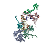
|
|---|---|
| 1 |
|
-Components
-RNA chain , 3 types, 4 molecules DCQR
| #1: RNA chain | Mass: 24890.121 Da / Num. of mol.: 2 / Source method: isolated from a natural source / Details: TAKEN FROM PDB ENTRIES 1GIX, 1TRA / Source: (natural) #2: RNA chain | | Mass: 13369.038 Da / Num. of mol.: 1 / Source method: isolated from a natural source / Details: TAKEN FROM PDB ENTRY 1GIY / Source: (natural) #3: RNA chain | | Mass: 8688.230 Da / Num. of mol.: 1 / Source method: isolated from a natural source / Details: TAKEN FROM PDB ENTRY 1GIY / Source: (natural) |
|---|
-Protein , 4 types, 4 molecules AOPL
| #4: Protein | Mass: 46064.723 Da / Num. of mol.: 1 / Source method: isolated from a natural source / Details: TAKEN FROM PDB ENTRY 1B23 / Source: (natural) |
|---|---|
| #5: Protein | Mass: 14920.754 Da / Num. of mol.: 1 / Source method: isolated from a natural source / Details: TAKEN FROM PDB ENTRY 1GIX / Source: (natural) |
| #6: Protein | Mass: 14338.861 Da / Num. of mol.: 1 / Source method: isolated from a natural source / Details: TAKEN FROM PDB ENTRY 1GIX / Source: (natural) |
| #7: Protein | Mass: 15111.923 Da / Num. of mol.: 1 / Source method: isolated from a natural source / Details: TAKEN FROM PDB ENTRY 1GIY / Source: (natural) |
-Experimental details
-Experiment
| Experiment | Method: ELECTRON MICROSCOPY |
|---|---|
| EM experiment | Aggregation state: PARTICLE / 3D reconstruction method: single particle reconstruction |
- Sample preparation
| Component | Name: EF-Tu/tRNA/GTP E. COLI 70S RIBOSOME / Type: RIBOSOME |
|---|---|
| Buffer solution | Name: Tris-HCl / pH: 7.5 / Details: Tris-HCl |
| Specimen | Embedding applied: NO / Shadowing applied: NO / Staining applied: NO / Vitrification applied: YES |
| Vitrification | Cryogen name: ETHANE |
| Crystal grow | *PLUS Method: cryo-electron microscopy / Details: cryo-electron microscopy |
- Electron microscopy imaging
| Microscopy | Model: FEI/PHILIPS CM200FEG/SOPHIE / Date: Mar 10, 2000 |
|---|---|
| Electron gun | Electron source:  FIELD EMISSION GUN / Accelerating voltage: 120 kV / Illumination mode: FLOOD BEAM FIELD EMISSION GUN / Accelerating voltage: 120 kV / Illumination mode: FLOOD BEAM |
| Electron lens | Mode: BRIGHT FIELD / Calibrated magnification: 58500 X / Nominal defocus max: 2500 nm / Nominal defocus min: 800 nm / Cs: 1.35 mm |
| Specimen holder | Temperature: 4.2 K / Tilt angle max: 0 ° / Tilt angle min: 0 ° |
| Image recording | Electron dose: 15 e/Å2 / Film or detector model: KODAK SO-163 FILM |
- Processing
| Software |
| ||||||||||||||||||||||||||||
|---|---|---|---|---|---|---|---|---|---|---|---|---|---|---|---|---|---|---|---|---|---|---|---|---|---|---|---|---|---|
| EM software |
| ||||||||||||||||||||||||||||
| CTF correction | Details: phase flip | ||||||||||||||||||||||||||||
| Symmetry | Point symmetry: C1 (asymmetric) | ||||||||||||||||||||||||||||
| 3D reconstruction | Method: "exact filter" backprojection / Resolution: 13 Å / Num. of particles: 24000 / Actual pixel size: 2.25 Å / Symmetry type: POINT | ||||||||||||||||||||||||||||
| Atomic model building | Protocol: RIGID BODY FIT / Space: REAL Target criteria: best visual fit using the program Amira (ribosomal proteins), best fit using the program Situs (EF-Tu) Details: REFINEMENT PROTOCOL--rigid body DETAILS--FOR S12, S13, SRL, HELIX69 ANS L11 ONLY BACKBONE COORDINATES ARE DEPOSITED | ||||||||||||||||||||||||||||
| Atomic model building |
| ||||||||||||||||||||||||||||
| Refinement step | Cycle: LAST
|