 Movie
Movie Controller

[English] 日本語
 Yorodumi
- EMDB-6306: Cryo-EM structure of the Bacillus subtilis MifM-stalled ribosome ... -
+ Open data

- Basic information
| Entry | Database: EMDB / ID: EMD-6306 | |||||||||
|---|---|---|---|---|---|---|---|---|---|---|
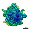
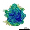
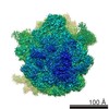
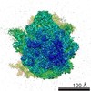
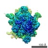
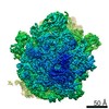
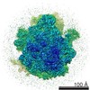
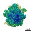
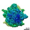
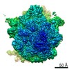
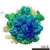
| Title | Cryo-EM structure of the Bacillus subtilis MifM-stalled ribosome complex | |||||||||
 Map data Map data | Cryo-EM structure of the B. subtilis MifM-stalled ribosome complex | |||||||||
 Sample Sample |
| |||||||||
 Keywords Keywords |  Bacillus subtilis / ribosome / cryo-EM / atomic model / stalling / translation arrest / MifM / L22 Bacillus subtilis / ribosome / cryo-EM / atomic model / stalling / translation arrest / MifM / L22 | |||||||||
| Function / homology |  Function and homology information Function and homology informationpositive regulation of rRNA processing / nucleoid / ribosomal small subunit biogenesis / small ribosomal subunit rRNA binding / ribosomal small subunit assembly / rRNA processing / cytosolic small ribosomal subunit / large ribosomal subunit rRNA binding / large ribosomal subunit / cytoplasmic translation ...positive regulation of rRNA processing / nucleoid / ribosomal small subunit biogenesis / small ribosomal subunit rRNA binding / ribosomal small subunit assembly / rRNA processing / cytosolic small ribosomal subunit / large ribosomal subunit rRNA binding / large ribosomal subunit / cytoplasmic translation / small ribosomal subunit / 5S rRNA binding / cytosolic large ribosomal subunit / transferase activity / tRNA binding / negative regulation of translation / membrane => GO:0016020 / rRNA binding / ribosome / structural constituent of ribosome / ribonucleoprotein complex / translation / response to antibiotic / mRNA binding / DNA binding / RNA binding / zinc ion binding / metal ion binding / plasma membrane / cytosol / cytoplasmSimilarity search - Function | |||||||||
| Biological species |  Bacillus subtilis (bacteria) Bacillus subtilis (bacteria) | |||||||||
| Method | single particle reconstruction / cryo EM / Resolution: 3.9 Å | |||||||||
 Authors Authors | Sohmen D / Chiba S / Shimokawa-Chiba N / Innis A / Berninghausen O / Beckmann R / Wilson DN | |||||||||
 Citation Citation | Journal: Nat Commun / Year: 2015 Title: Structure of the Bacillus subtilis 70S ribosome reveals the basis for species-specific stalling. Authors: Daniel Sohmen / Shinobu Chiba / Naomi Shimokawa-Chiba / C Axel Innis / Otto Berninghausen / Roland Beckmann / Koreaki Ito / Daniel N Wilson /    Abstract: Ribosomal stalling is used to regulate gene expression and can occur in a species-specific manner. Stalling during translation of the MifM leader peptide regulates expression of the downstream ...Ribosomal stalling is used to regulate gene expression and can occur in a species-specific manner. Stalling during translation of the MifM leader peptide regulates expression of the downstream membrane protein biogenesis factor YidC2 (YqjG) in Bacillus subtilis, but not in Escherichia coli. In the absence of structures of Gram-positive bacterial ribosomes, a molecular basis for species-specific stalling has remained unclear. Here we present the structure of a Gram-positive B. subtilis MifM-stalled 70S ribosome at 3.5-3.9 Å, revealing a network of interactions between MifM and the ribosomal tunnel, which stabilize a non-productive conformation of the PTC that prevents aminoacyl-tRNA accommodation and thereby induces translational arrest. Complementary genetic analyses identify a single amino acid within ribosomal protein L22 that dictates the species specificity of the stalling event. Such insights expand our understanding of how the synergism between the ribosome and the nascent chain is utilized to modulate the translatome in a species-specific manner. | |||||||||
| History |
|
- Structure visualization
| Movie |
Movie viewer |
|---|---|
| Structure viewer | EM map: SurfViewMolmilJmol/JSmol |
| Supplemental images |

- Downloads & links
-EMDB archive
| Map data | emd_6306.map.gz | 173 MB | EMDB map data format | |
|---|---|---|---|---|
| Header (meta data) | emd-6306-v30.xmlemd-6306.xml | 9.9 KB 9.9 KB | Display Display | EMDB header |
| Images | 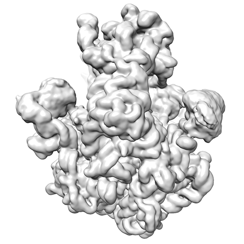 emd_6306.jpg emd_6306.jpg | 107.1 KB | ||
| Archive directory |  http://ftp.pdbj.org/pub/emdb/structures/EMD-6306ftp://ftp.pdbj.org/pub/emdb/structures/EMD-6306 http://ftp.pdbj.org/pub/emdb/structures/EMD-6306ftp://ftp.pdbj.org/pub/emdb/structures/EMD-6306 | HTTPS FTP |
-Related structure data
| Related structure data |  3j9wMC M: atomic model generated by this map C: citing same article ( |
|---|---|
| Similar structure data |
-Links
| EMDB pages | EMDB (EBI/PDBe) / EMDataResource |
|---|---|
| Related items in Molecule of the Month |


-Map
| File | Download / File: emd_6306.map.gz / Format: CCP4 / Size: 185.7 MB / Type: IMAGE STORED AS FLOATING POINT NUMBER (4 BYTES) | ||||||||||||||||||||||||||||||||||||||||||||||||||||||||||||
|---|---|---|---|---|---|---|---|---|---|---|---|---|---|---|---|---|---|---|---|---|---|---|---|---|---|---|---|---|---|---|---|---|---|---|---|---|---|---|---|---|---|---|---|---|---|---|---|---|---|---|---|---|---|---|---|---|---|---|---|---|---|
| Annotation | Cryo-EM structure of the B. subtilis MifM-stalled ribosome complex | ||||||||||||||||||||||||||||||||||||||||||||||||||||||||||||
| Voxel size | X=Y=Z: 1.108 Å | ||||||||||||||||||||||||||||||||||||||||||||||||||||||||||||
| Density |
| ||||||||||||||||||||||||||||||||||||||||||||||||||||||||||||
| Symmetry | Space group: 1 | ||||||||||||||||||||||||||||||||||||||||||||||||||||||||||||
| Details | EMDB XML:
CCP4 map header:
| ||||||||||||||||||||||||||||||||||||||||||||||||||||||||||||
-Supplemental data
- Sample components
-Entire : B. subtilis MifM-stalled ribosome complex
| Entire | Name: B. subtilis MifM-stalled ribosome complex |
|---|---|
| Components |
|
-Supramolecule #1000: B. subtilis MifM-stalled ribosome complex
| Supramolecule | Name: B. subtilis MifM-stalled ribosome complex / type: sample / ID: 1000 / Number unique components: 3 |
|---|
-Supramolecule #1: 70S ribosome
| Supramolecule | Name: 70S ribosome / type: complex / ID: 1 / Details: stalled / Recombinant expression: No / Database: NCBI Ribosome-details: ribosome-prokaryote: LSU 50S, LSU RNA 23S, LSU RNA 5S, SSU 30S, PSR16s, ALL |
|---|---|
| Source (natural) | Organism: Bacillus subtilis (bacteria) / Strain: 168 |
-Macromolecule #1: Membrane protein insertion and folding monitor
| Macromolecule | Name: Membrane protein insertion and folding monitor / type: protein_or_peptide / ID: 1 / Name.synonym: MifM / Number of copies: 1 / Oligomeric state: monomer / Recombinant expression: No / Database: NCBI |
|---|---|
| Source (natural) | Organism: Bacillus subtilis (bacteria) / Strain: 168 |
-Macromolecule #2: tRNA-Asp
| Macromolecule | Name: tRNA-Asp / type: rna / ID: 2 / Classification: TRANSFER / Structure: DOUBLE HELIX / Synthetic?: No |
|---|---|
| Source (natural) | Organism: Bacillus subtilis (bacteria) / Strain: 168 |
-Experimental details
-Structure determination
| Method | cryo EM |
|---|---|
 Processing Processing | single particle reconstruction |
| Aggregation state | particle |
-Sample preparation
| Buffer | pH: 7.2 |
|---|---|
| Grid | Details: 2 nm pre-coated Quantifoil R3/3 holey carbon support grids |
| Vitrification | Cryogen name: ETHANE / Instrument: FEI VITROBOT MARK IV |
- Electron microscopy
| Microscope | FEI TITAN KRIOS |
|---|---|
| Electron beam | Acceleration voltage: 300 kV / Electron source: FIELD EMISSION GUN |
| Electron optics | Illumination mode: SPOT SCAN / Imaging mode: BRIGHT FIELDBright-field microscopy / Nominal defocus max: 2.5 µm / Nominal defocus min: 1.0 µm / Nominal magnification: 125085 / Cs: mm |
| Sample stage | Specimen holder model: FEI TITAN KRIOS AUTOGRID HOLDER |
| Cs | 0 |
| Date | Apr 12, 2014 |
| Image recording | Category: CCD / Film or detector model: FEI FALCON II (4k x 4k) / Average electron dose: 28 e/Å2 |
| Experimental equipment |  Model: Titan Krios / Image courtesy: FEI Company |
-Image processing
| CTF correction | Details: defocus groups |
|---|---|
| Final reconstruction | Resolution.type: BY AUTHOR / Resolution: 3.9 Å / Resolution method: OTHER / Software - Name: SPIDER Details: Since images from microscopy were processed in the absence of spatial frequencies higher than 8 A, a FSC cut-off value of 0.143 was used for average resolution determination of 3.9 A (Scheres and Chen, 2012). Number images used: 305045 |
