 Movie
Movie Controller

[English] 日本語
 Yorodumi
- EMDB-3396: Cryo-electron microscopy structure of the star-shaped, hubless po... -
+ Open data

- Basic information
| Entry | Database: EMDB / ID: EMD-3396 | |||||||||
|---|---|---|---|---|---|---|---|---|---|---|
| Title | Cryo-electron microscopy structure of the star-shaped, hubless post-attachment T4 baseplate | |||||||||
 Map data Map data | Cryo-electron microscopy structure of the star-shaped, hubless post-attachment T4 baseplate. Postprocessed with Relion, using automated B-factor estimation and the mask used for polishing, which was generated previously by auto-masking with an initial binarization threshold of 0.015. | |||||||||
 Sample Sample |
| |||||||||
 Keywords Keywords | T4 /  baseplate / post-attachment / bacteriophage / bacterial virus / star-shaped / hubless / membrane-piercing / cell attachment / infection baseplate / post-attachment / bacteriophage / bacterial virus / star-shaped / hubless / membrane-piercing / cell attachment / infection | |||||||||
| Function / homology |  Function and homology information Function and homology informationvirus tail, baseplate / viral tail assembly / viral release from host cell / identical protein binding Similarity search - Function | |||||||||
| Biological species |  Enterobacteria phage T4 (virus) Enterobacteria phage T4 (virus) | |||||||||
| Method | single particle reconstruction / cryo EM / Resolution: 6.77 Å | |||||||||
 Authors Authors | Taylor NMI / Guerrero-Ferreira RC / Goldie KN / Stahlberg H / Leiman PG | |||||||||
 Citation Citation | Journal: Nature / Year: 2016 Title: Structure of the T4 baseplate and its function in triggering sheath contraction. Authors: Nicholas M I Taylor / Nikolai S Prokhorov / Ricardo C Guerrero-Ferreira / Mikhail M Shneider / Christopher Browning / Kenneth N Goldie / Henning Stahlberg / Petr G Leiman /   Abstract: Several systems, including contractile tail bacteriophages, the type VI secretion system and R-type pyocins, use a multiprotein tubular apparatus to attach to and penetrate host cell membranes. This ...Several systems, including contractile tail bacteriophages, the type VI secretion system and R-type pyocins, use a multiprotein tubular apparatus to attach to and penetrate host cell membranes. This macromolecular machine resembles a stretched, coiled spring (or sheath) wound around a rigid tube with a spike-shaped protein at its tip. A baseplate structure, which is arguably the most complex part of this assembly, relays the contraction signal to the sheath. Here we present the atomic structure of the approximately 6-megadalton bacteriophage T4 baseplate in its pre- and post-host attachment states and explain the events that lead to sheath contraction in atomic detail. We establish the identity and function of a minimal set of components that is conserved in all contractile injection systems and show that the triggering mechanism is universally conserved. | |||||||||
| History |
|
- Structure visualization
| Movie |
Movie viewer |
|---|---|
| Structure viewer | EM map: SurfViewMolmilJmol/JSmol |
| Supplemental images |

- Downloads & links
-EMDB archive
| Map data | emd_3396.map.gz | 28.3 MB | EMDB map data format | |
|---|---|---|---|---|
| Header (meta data) | emd-3396-v30.xmlemd-3396.xml | 10.6 KB 10.6 KB | Display Display | EMDB header |
| FSC (resolution estimation) | emd_3396_fsc.xml | 16.5 KB | Display | FSC data file |
| Images | 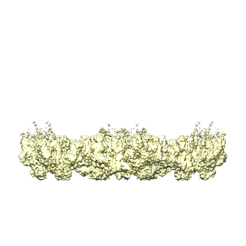 emd_3396.png emd_3396.png | 99.3 KB | ||
| Archive directory |  http://ftp.pdbj.org/pub/emdb/structures/EMD-3396ftp://ftp.pdbj.org/pub/emdb/structures/EMD-3396 http://ftp.pdbj.org/pub/emdb/structures/EMD-3396ftp://ftp.pdbj.org/pub/emdb/structures/EMD-3396 | HTTPS FTP |
-Related structure data
| Related structure data |  5iv7MC 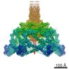 3374C 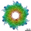 3392C  3393C  3394C 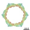 3395C  3397C  5iv5C  5iw9C M: atomic model generated by this map C: citing same article ( |
|---|---|
| Similar structure data |
-Links
| EMDB pages | EMDB (EBI/PDBe) / EMDataResource |
|---|
-Map
| File | Download / File: emd_3396.map.gz / Format: CCP4 / Size: 412 MB / Type: IMAGE STORED AS FLOATING POINT NUMBER (4 BYTES) | ||||||||||||||||||||||||||||||||||||||||||||||||||||||||||||
|---|---|---|---|---|---|---|---|---|---|---|---|---|---|---|---|---|---|---|---|---|---|---|---|---|---|---|---|---|---|---|---|---|---|---|---|---|---|---|---|---|---|---|---|---|---|---|---|---|---|---|---|---|---|---|---|---|---|---|---|---|---|
| Annotation | Cryo-electron microscopy structure of the star-shaped, hubless post-attachment T4 baseplate. Postprocessed with Relion, using automated B-factor estimation and the mask used for polishing, which was generated previously by auto-masking with an initial binarization threshold of 0.015. | ||||||||||||||||||||||||||||||||||||||||||||||||||||||||||||
| Voxel size | X=Y=Z: 1.326 Å | ||||||||||||||||||||||||||||||||||||||||||||||||||||||||||||
| Density |
| ||||||||||||||||||||||||||||||||||||||||||||||||||||||||||||
| Symmetry | Space group: 1 | ||||||||||||||||||||||||||||||||||||||||||||||||||||||||||||
| Details | EMDB XML:
CCP4 map header:
| ||||||||||||||||||||||||||||||||||||||||||||||||||||||||||||
-Supplemental data
- Sample components
-Entire : Star-shaped, hubless post-attachment T4 baseplate
| Entire | Name: Star-shaped, hubless post-attachment T4 baseplate |
|---|---|
| Components |
|
-Supramolecule #1000: Star-shaped, hubless post-attachment T4 baseplate
| Supramolecule | Name: Star-shaped, hubless post-attachment T4 baseplate / type: sample / ID: 1000 Details: In addition to hexagonal pre-attachment baseplate-tail tube complexes, the sample also contained some star-shaped, hubless post-attachment baseplates. The current reconstruction is the ...Details: In addition to hexagonal pre-attachment baseplate-tail tube complexes, the sample also contained some star-shaped, hubless post-attachment baseplates. The current reconstruction is the reconstruction of those post-attachment, star-shaped baseplates, which have the following oligomeric state: (gp6)12(gp7)6(gp8)12(gp9)18(gp10)18(gp11)18(gp12)18(gp25)6(gp53)6. This oligomeric state has an approximate theoretical MW of 5.5 MDa. Oligomeric state: (gp6)12(gp7)6(gp8)12(gp9)18(gp10)18(gp11)18(gp12)18(gp25)6(gp53)6 Number unique components: 1 |
|---|---|
| Molecular weight | Theoretical: 5.5 MDa |
-Macromolecule #1: Star-shaped, hubless post-attachment T4 baseplate
| Macromolecule | Name: Star-shaped, hubless post-attachment T4 baseplate / type: protein_or_peptide / ID: 1 / Number of copies: 1 / Recombinant expression: No |
|---|---|
| Source (natural) | Organism: Enterobacteria phage T4 (virus) / Strain: am18/am23 mutant |
| Molecular weight | Theoretical: 8.7 MDa |
-Experimental details
-Structure determination
| Method | cryo EM |
|---|---|
 Processing Processing | single particle reconstruction |
| Aggregation state | particle |
-Sample preparation
| Concentration | 1 mg/mL |
|---|---|
| Buffer | pH: 8 / Details: 50 mM Tris-HCl pH 8.0, 100 mM NaCl, 8 mM MgSO4 |
| Grid | Details: Quantifoil 300 mesh carbon-coated copper grids glow-discharged for 20 seconds |
| Vitrification | Cryogen name: ETHANE / Chamber humidity: 100 % / Instrument: FEI VITROBOT MARK IV Method: Applied 3.5 ul of sample and blotting 3 seconds before plunging |
- Electron microscopy
| Microscope | FEI TITAN KRIOS |
|---|---|
| Electron beam | Acceleration voltage: 300 kV / Electron source: FIELD EMISSION GUN |
| Electron optics | Calibrated magnification: 37700 / Illumination mode: FLOOD BEAM / Imaging mode: BRIGHT FIELDBright-field microscopy / Cs: 2.7 mm / Nominal defocus max: 4.0 µm / Nominal defocus min: 0.5 µm / Nominal magnification: 105000 |
| Specialist optics | Energy filter - Name: Quantum-LS Gatan Image Filter / Energy filter - Lower energy threshold: 0.0 eV / Energy filter - Upper energy threshold: 20.0 eV |
| Sample stage | Specimen holder model: FEI TITAN KRIOS AUTOGRID HOLDER |
| Temperature | Average: 80 K |
| Alignment procedure | Legacy - Astigmatism: The astigmatism was corrected at high magnification |
| Date | May 7, 2015 |
| Image recording | Category: CCD / Film or detector model: GATAN K2 SUMMIT (4k x 4k) / Number real images: 1621 / Average electron dose: 60 e/Å2 Details: Individual frames were aligned with 2dx_automator. 40 frames were recorded in total, and the 2 first frames were discarded. Bits/pixel: 32 |
| Experimental equipment |  Model: Titan Krios / Image courtesy: FEI Company |
-Image processing
| CTF correction | Details: Each particle |
|---|---|
| Final reconstruction | Applied symmetry - Point group: C6 (6 fold cyclic) / Algorithm: OTHER / Resolution.type: BY AUTHOR / Resolution: 6.77 Å / Resolution method: OTHER / Software - Name: RELION Details: The short tail fibers (gp12 trimers) appear more flexible, and no special effort was made to reconstruct them. They were partly masked out by the circular mask.The total mass of the ...Details: The short tail fibers (gp12 trimers) appear more flexible, and no special effort was made to reconstruct them. They were partly masked out by the circular mask.The total mass of the reconstructed volume was approximately 4.5 MDa. Number images used: 5176 |
| Details | The particles were selected with e2boxer.py. |
| FSC plot (resolution estimation) |  |
