Movie
Movie Controller


+ Open data

- Basic information
| Entry | Database: EMDB / ID: EMD-2554 | |||||||||
|---|---|---|---|---|---|---|---|---|---|---|
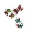
| Title | electron tomography average of an IgG hexamer | |||||||||
 Map data Map data | reconstruction of IgG1-005-RGY hexamer | |||||||||
 Sample Sample |
| |||||||||
 Keywords Keywords | IgG hexamer / IgG / classical complement activation | |||||||||
| Biological species |  Homo sapiens (human) Homo sapiens (human) | |||||||||
| Method | subtomogram averaging / negative staining / Resolution: 22.0 Å | |||||||||
 Authors Authors | Diebolder CA / Beurskens FJ / de Jong RN / Koning RI / Strumane K / Lindorfer MA / Voorhorst M / Ugurlar D / Rosati S / Heck AJR ...Diebolder CA / Beurskens FJ / de Jong RN / Koning RI / Strumane K / Lindorfer MA / Voorhorst M / Ugurlar D / Rosati S / Heck AJR / van de Winkel JGJ / Wilson IA / Koster AJ / Taylor RP / Ollmann-Saphire E / Burton DR / Schuurman J / Gros P / Parren PWHI | |||||||||
 Citation Citation | Journal: Science / Year: 2014 Title: Complement is activated by IgG hexamers assembled at the cell surface. Authors: Christoph A Diebolder / Frank J Beurskens / Rob N de Jong / Roman I Koning / Kristin Strumane / Margaret A Lindorfer / Marleen Voorhorst / Deniz Ugurlar / Sara Rosati / Albert J R Heck / Jan ...Authors: Christoph A Diebolder / Frank J Beurskens / Rob N de Jong / Roman I Koning / Kristin Strumane / Margaret A Lindorfer / Marleen Voorhorst / Deniz Ugurlar / Sara Rosati / Albert J R Heck / Jan G J van de Winkel / Ian A Wilson / Abraham J Koster / Ronald P Taylor / Erica Ollmann Saphire / Dennis R Burton / Janine Schuurman / Piet Gros / Paul W H I Parren /  Abstract: Complement activation by antibodies bound to pathogens, tumors, and self antigens is a critical feature of natural immune defense, a number of disease processes, and immunotherapies. How antibodies ...Complement activation by antibodies bound to pathogens, tumors, and self antigens is a critical feature of natural immune defense, a number of disease processes, and immunotherapies. How antibodies activate the complement cascade, however, is poorly understood. We found that specific noncovalent interactions between Fc segments of immunoglobulin G (IgG) antibodies resulted in the formation of ordered antibody hexamers after antigen binding on cells. These hexamers recruited and activated C1, the first component of complement, thereby triggering the complement cascade. The interactions between neighboring Fc segments could be manipulated to block, reconstitute, and enhance complement activation and killing of target cells, using all four human IgG subclasses. We offer a general model for understanding antibody-mediated complement activation and the design of antibody therapeutics with enhanced efficacy. | |||||||||
| History |
|
- Structure visualization
| Movie |
 Movie viewer Movie viewer |
|---|---|
| Structure viewer | EM map: SurfViewMolmilJmol/JSmol |
| Supplemental images |

- Downloads & links
-EMDB archive
| Map data | emd_2554.map.gz | 14.8 MB | EMDB map data format | |
|---|---|---|---|---|
| Header (meta data) | emd-2554-v30.xmlemd-2554.xml | 12.7 KB 12.7 KB | Display Display | EMDB header |
| Images | 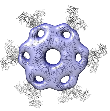 emd_2554.png emd_2554.png | 188.9 KB | ||
| Masks | emd_2554_msk_1.map | 20.1 MB | Mask map | |
| Archive directory |  http://ftp.pdbj.org/pub/emdb/structures/EMD-2554ftp://ftp.pdbj.org/pub/emdb/structures/EMD-2554 http://ftp.pdbj.org/pub/emdb/structures/EMD-2554ftp://ftp.pdbj.org/pub/emdb/structures/EMD-2554 | HTTPS FTP |
-Validation report
| Summary document | emd_2554_validation.pdf.gz | 229.7 KB | Display | EMDB validaton report |
|---|---|---|---|---|
| Full document | emd_2554_full_validation.pdf.gz | 228.7 KB | Display | |
| Data in XML | emd_2554_validation.xml.gz | 6.1 KB | Display | |
| Arichive directory | https://ftp.pdbj.org/pub/emdb/validation_reports/EMD-2554ftp://ftp.pdbj.org/pub/emdb/validation_reports/EMD-2554 | HTTPS FTP |
-Related structure data
-Links
| EMDB pages | EMDB (EBI/PDBe) / EMDataResource |
|---|
-Map
| File | Download / File: emd_2554.map.gz / Format: CCP4 / Size: 19.6 MB / Type: IMAGE STORED AS FLOATING POINT NUMBER (4 BYTES) | ||||||||||||||||||||||||||||||||||||||||||||||||||||||||||||||||||||
|---|---|---|---|---|---|---|---|---|---|---|---|---|---|---|---|---|---|---|---|---|---|---|---|---|---|---|---|---|---|---|---|---|---|---|---|---|---|---|---|---|---|---|---|---|---|---|---|---|---|---|---|---|---|---|---|---|---|---|---|---|---|---|---|---|---|---|---|---|---|
| Annotation | reconstruction of IgG1-005-RGY hexamer | ||||||||||||||||||||||||||||||||||||||||||||||||||||||||||||||||||||
| Voxel size | X=Y=Z: 4.4 Å | ||||||||||||||||||||||||||||||||||||||||||||||||||||||||||||||||||||
| Density |
| ||||||||||||||||||||||||||||||||||||||||||||||||||||||||||||||||||||
| Symmetry | Space group: 1 | ||||||||||||||||||||||||||||||||||||||||||||||||||||||||||||||||||||
| Details | EMDB XML:
CCP4 map header:
| ||||||||||||||||||||||||||||||||||||||||||||||||||||||||||||||||||||
-Supplemental data
-Segmentation: this mask represents the FC hexamer
| Annotation | this mask represents the FC hexamer | ||||||||||||
|---|---|---|---|---|---|---|---|---|---|---|---|---|---|
| File | emd_2554_msk_1.map | ||||||||||||
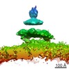
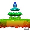
| Projections & Slices |
| ||||||||||||
| Density Histograms |
 Z
Z Y
Y X
X


- Sample components
-Entire : IgG1-005-RGY monoclonal antibody
| Entire | Name: IgG1-005-RGY monoclonal antibody |
|---|---|
| Components |
|
-Supramolecule #1000: IgG1-005-RGY monoclonal antibody
| Supramolecule | Name: IgG1-005-RGY monoclonal antibody / type: sample / ID: 1000 / Details: sample is a mix of monomers and hexamers / Oligomeric state: one homohexamer / Number unique components: 1 |
|---|---|
| Molecular weight | Experimental: 890 KDa / Method: native mass spectrometry |
-Macromolecule #1: IgG1-005-E345R-E430G-S440Y
| Macromolecule | Name: IgG1-005-E345R-E430G-S440Y / type: protein_or_peptide / ID: 1 / Number of copies: 6 / Oligomeric state: Hexamer / Recombinant expression: Yes |
|---|---|
| Source (natural) | Organism: Homo sapiens (human) / synonym: Human |
| Molecular weight | Experimental: 148.5 KDa / Theoretical: 145.4 KDa |
| Recombinant expression | Organism: Homo sapiens (human) / Recombinant cell: Freestyle 293-F / Recombinant plasmid: pcDNA3.3 |
-Experimental details
-Structure determination
| Method | negative staining |
|---|---|
 Processing Processing | subtomogram averaging |
| Aggregation state | particle |
-Sample preparation
| Concentration | 0.03 mg/mL |
|---|---|
| Staining | Type: NEGATIVE / Details: negative staining with 3% uranyl acetate |
| Grid | Details: 400 mesh copper grids with carbon support |
| Vitrification | Cryogen name: NONE / Instrument: OTHER |
- Electron microscopy
| Microscope | FEI TECNAI F20 |
|---|---|
| Alignment procedure | Legacy - Astigmatism: Objective lens astigmatism was corrected at 50,000 times magnification |
| Date | Sep 3, 2013 |
| Image recording | Category: CCD / Film or detector model: GATAN ULTRASCAN 4000 (4k x 4k) |
| Electron beam | Acceleration voltage: 200 kV / Electron source:  FIELD EMISSION GUN FIELD EMISSION GUN |
| Electron optics | Calibrated magnification: 50000 / Illumination mode: FLOOD BEAM / Imaging mode: BRIGHT FIELD / Cs: 2 mm / Nominal defocus max: 1.035 µm / Nominal defocus min: 1.01 µm / Nominal magnification: 50000 |
| Sample stage | Specimen holder model: SIDE ENTRY, EUCENTRIC / Tilt series - Axis1 - Min angle: -66 ° / Tilt series - Axis1 - Max angle: 66 ° |
| Experimental equipment |  Model: Tecnai F20 / Image courtesy: FEI Company |
-Image processing
| Details | manual picking of 237 particles from two tomograms (PEET stalkInit), random particle as initial reference, iterative refinement in PEET |
|---|---|
| Final reconstruction | Applied symmetry - Point group: C1 (asymmetric) / Algorithm: OTHER / Resolution.type: BY AUTHOR / Resolution: 22.0 Å / Resolution method: OTHER / Software - Name: IMOD, PEET, TOMOCTF / Details: final map was calculated from 200 sub tomograms / Number subtomograms used: 200 |
-Atomic model buiding 1
| Initial model | PDB ID: |
|---|---|
| Software | Name: Chimera |
| Details | manual docking |
| Refinement | Space: REAL / Protocol: RIGID BODY FIT |