 Movie
Movie Controller

[English] 日本語
 Yorodumi
- EMDB-2061: Icosahedral reconstruction of Salisaeta icosahedral phage 1 (SSIP-1) -
+ Open data

- Basic information
| Entry | Database: EMDB / ID: EMD-2061 | |||||||||
|---|---|---|---|---|---|---|---|---|---|---|
| Title | Icosahedral reconstruction of Salisaeta icosahedral phage 1 (SSIP-1) | |||||||||
 Map data Map data | Icosahedral reconstruction of phage SSIP-1 | |||||||||
 Sample Sample |
| |||||||||
 Keywords Keywords | Salisaeta icosahedral phage 1 / SSIP-1 / SSIP1 / halophilic /  phage / bacteriophage / virus phage / bacteriophage / virus | |||||||||
| Biological species |  Salisaeta icosahedral phage 1 (virus) Salisaeta icosahedral phage 1 (virus) | |||||||||
| Method | single particle reconstruction / cryo EM / Resolution: 12.5 Å | |||||||||
 Authors Authors | Aalto AP / Bitto D / Ravantti JJ / Bamford DH / Huiskonen JT / Oksanen HM | |||||||||
 Citation Citation | Journal: Proc Natl Acad Sci U S A / Year: 2012 Title: Snapshot of virus evolution in hypersaline environments from the characterization of a membrane-containing Salisaeta icosahedral phage 1. Authors: Antti P Aalto / David Bitto / Janne J Ravantti / Dennis H Bamford / Juha T Huiskonen / Hanna M Oksanen /  Abstract: The multitude of archaea and bacteria inhabiting extreme environments has only become evident during the last decades. As viruses apply a significant evolutionary force to their hosts, there is an ...The multitude of archaea and bacteria inhabiting extreme environments has only become evident during the last decades. As viruses apply a significant evolutionary force to their hosts, there is an inherent value in learning about viruses infecting these extremophiles. In this study, we have focused on one such unique virus-host pair isolated from a hypersaline environment: an icosahedral, membrane-containing double-stranded DNA virus--Salisaeta icosahedral phage 1 (SSIP-1) and its halophilic host bacterium Salisaeta sp. SP9-1 closely related to Salisaeta longa. The architectural principles, virion composition, and the proposed functions associated with some of the ORFs of the virus are surprisingly similar to those found in viruses belonging to the PRD1-adenovirus lineage. The virion structure, determined by electron cryomicroscopy, reveals that the bulk of the outer protein capsid is composed of upright standing pseudohexameric capsomers organized on a T = 49 icosahedral lattice. Our results give a comprehensive description of a halophilic virus-host system and shed light on the relatedness of viruses based on their virion architecture. | |||||||||
| History |
|
- Structure visualization
| Movie |
 Movie viewer Movie viewer |
|---|---|
| Structure viewer | EM map: SurfViewMolmilJmol/JSmol |
| Supplemental images |

- Downloads & links
-EMDB archive
| Map data | emd_2061.map.gz | 455.6 MB | EMDB map data format | |
|---|---|---|---|---|
| Header (meta data) | emd-2061-v30.xmlemd-2061.xml | 9 KB 9 KB | Display Display | EMDB header |
| Images | 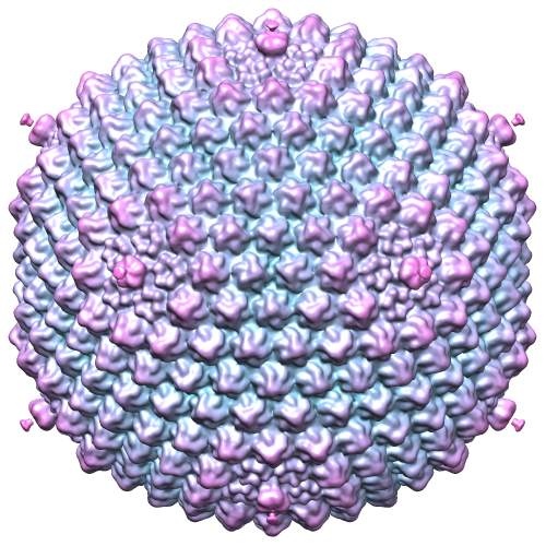 emd_2061.png emd_2061.png | 390.2 KB | ||
| Archive directory |  http://ftp.pdbj.org/pub/emdb/structures/EMD-2061ftp://ftp.pdbj.org/pub/emdb/structures/EMD-2061 http://ftp.pdbj.org/pub/emdb/structures/EMD-2061ftp://ftp.pdbj.org/pub/emdb/structures/EMD-2061 | HTTPS FTP |
-Related structure data
| Similar structure data |
|---|
-Links
| EMDB pages | EMDB (EBI/PDBe) / EMDataResource |
|---|
-Map
| File | Download / File: emd_2061.map.gz / Format: CCP4 / Size: 1.4 GB / Type: IMAGE STORED AS FLOATING POINT NUMBER (4 BYTES) | ||||||||||||||||||||||||||||||||||||||||||||||||||||||||||||||||||||
|---|---|---|---|---|---|---|---|---|---|---|---|---|---|---|---|---|---|---|---|---|---|---|---|---|---|---|---|---|---|---|---|---|---|---|---|---|---|---|---|---|---|---|---|---|---|---|---|---|---|---|---|---|---|---|---|---|---|---|---|---|---|---|---|---|---|---|---|---|---|
| Annotation | Icosahedral reconstruction of phage SSIP-1 | ||||||||||||||||||||||||||||||||||||||||||||||||||||||||||||||||||||
| Voxel size | X=Y=Z: 2 Å | ||||||||||||||||||||||||||||||||||||||||||||||||||||||||||||||||||||
| Density |
| ||||||||||||||||||||||||||||||||||||||||||||||||||||||||||||||||||||
| Symmetry | Space group: 1 | ||||||||||||||||||||||||||||||||||||||||||||||||||||||||||||||||||||
| Details | EMDB XML:
CCP4 map header:
| ||||||||||||||||||||||||||||||||||||||||||||||||||||||||||||||||||||
-Supplemental data
- Sample components
-Entire : Salisaeta icosahedral phage 1 (SSIP1) virion
| Entire | Name: Salisaeta icosahedral phage 1 (SSIP1) virion |
|---|---|
| Components |
|
-Supramolecule #1000: Salisaeta icosahedral phage 1 (SSIP1) virion
| Supramolecule | Name: Salisaeta icosahedral phage 1 (SSIP1) virion / type: sample / ID: 1000 / Oligomeric state: Whole virion / Number unique components: 1 |
|---|
-Supramolecule #1: Salisaeta icosahedral phage 1
| Supramolecule | Name: Salisaeta icosahedral phage 1 / type: virus / ID: 1 / Name.synonym: SSIP1 / NCBI-ID: 1183239 / Sci species name: Salisaeta icosahedral phage 1 / Virus type: VIRION / Virus isolate: SPECIES / Virus enveloped: Yes / Virus empty: No / Syn species name: SSIP1 |
|---|---|
| Host (natural) | Organism:  Salisaeta sp. SP9-1 (bacteria) / synonym: BACTERIA(EUBACTERIA) Salisaeta sp. SP9-1 (bacteria) / synonym: BACTERIA(EUBACTERIA) |
| Virus shell | Shell ID: 1 / Name: Capsid / Diameter: 1000 Å / T number (triangulation number): 49 |
-Experimental details
-Structure determination
| Method | cryo EM |
|---|---|
 Processing Processing | single particle reconstruction |
| Aggregation state | particle |
-Sample preparation
| Buffer | Details: 9% salt water (SW) see http://www.haloarchaea.com/resources/halohandbook/Halohandbook_2008_v7.pdf |
|---|---|
| Grid | Details: 200 mesh molybdenum grid with holey carbon, glow discharged |
| Vitrification | Cryogen name: HELIUM / Chamber humidity: 80 % / Chamber temperature: 110 K / Instrument: GATAN CRYOPLUNGE 3 / Method: Blot for 4 seconds from opposite side |
- Electron microscopy
| Microscope | FEI POLARA 300 |
|---|---|
| Electron beam | Acceleration voltage: 300 kV / Electron source: FIELD EMISSION GUN |
| Electron optics | Calibrated magnification: 75000 / Illumination mode: FLOOD BEAM / Imaging mode: BRIGHT FIELDBright-field microscopy / Cs: 2 mm / Nominal defocus max: 2.6 µm / Nominal defocus min: 1.0 µm / Nominal magnification: 59000 |
| Sample stage | Specimen holder: liquid nitrogen temperature / Specimen holder model: OTHER |
| Temperature | Average: 81 K |
| Date | Feb 26, 2011 |
| Image recording | Category: CCD / Film or detector model: GATAN ULTRASCAN 4000 (4k x 4k) / Number real images: 842 / Average electron dose: 20 e/Å2 |
| Experimental equipment |  Model: Tecnai Polara / Image courtesy: FEI Company |
-Image processing
| CTF correction | Details: Each particle |
|---|---|
| Final reconstruction | Applied symmetry - Point group: I (icosahedral) / Algorithm: OTHER / Resolution.type: BY AUTHOR / Resolution: 12.5 Å / Resolution method: FSC 0.5 CUT-OFF / Software - Name: EMAN2 / Number images used: 2747 |
| Details | The particles were manually picked in EMAN2. Initial model was calculated in IMAGIC5. Contrast transfer function correction and icosahedral reconstruction was carried out in EMAN. The final map was low pass filtered to 11 A. |
