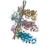
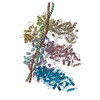
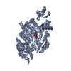
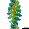
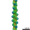
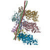
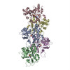
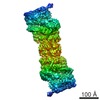
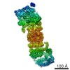
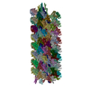
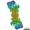
Journal: Cell / Year: 2012 Title: Structure of the rigor actin-tropomyosin-myosin complex. Authors: Elmar Behrmann / Mirco Müller / Pawel A Penczek / Hans Georg Mannherz / Dietmar J Manstein / Stefan Raunser / Abstract: Regulation of myosin and filamentous actin interaction by tropomyosin is a central feature of contractile events in muscle and nonmuscle cells. However, little is known about molecular interactions ...Regulation of myosin and filamentous actin interaction by tropomyosin is a central feature of contractile events in muscle and nonmuscle cells. However, little is known about molecular interactions within the complex and the trajectory of tropomyosin movement between its "open" and "closed" positions on the actin filament. Here, we report the 8 Å resolution structure of the rigor (nucleotide-free) actin-tropomyosin-myosin complex determined by cryo-electron microscopy. The pseudoatomic model of the complex, obtained from fitting crystal structures into the map, defines the large interface involving two adjacent actin monomers and one tropomyosin pseudorepeat per myosin contact. Severe forms of hereditary myopathies are linked to mutations that critically perturb this interface. Myosin binding results in a 23 Å shift of tropomyosin along actin. Complex domain motions occur in myosin, but not in actin. Based on our results, we propose a structural model for the tropomyosin-dependent modulation of myosin binding to actin.
History
Deposition
Nov 14, 2011
-
Header (metadata) release
Aug 1, 2012
-
Map release
Aug 1, 2012
-
Update
Aug 1, 2012
-
Current status
Aug 1, 2012
Processing site: PDBe / Status: Released
-
Structure visualization
Movie
Surface view with section colored by density value
Legacy - Astigmatism: objective lens astigmatism was corrected at 150,000 times magnification
Specialist optics
Energy filter - Name: in-column Omega filter / Energy filter - Lower energy threshold: 0.0 eV / Energy filter - Upper energy threshold: 12.0 eV
Image recording
Category: CCD / Film or detector model: TVIPS TEMCAM-F816 (8k x 8k) / Digitization - Sampling interval: 15.6 µm / Number real images: 836 / Average electron dose: 17 e/Å2 Details: Over 3000 images were taken of which only the best 836 were used for processing Bits/pixel: 14
Electron beam
Acceleration voltage: 200 kV / Electron source: FIELD EMISSION GUN
PDBEntryID_givenInChain. Protocol: geometry-based conformational sampling using Deformable Elastic Network (DEN) approach. Initial placement was performed using rigid-body fitting in Chimera
Refinement
Space: REAL / Protocol: FLEXIBLE FIT
Output model
PDB-4a7f: Structure of the Actin-Tropomyosin-Myosin Complex (rigor ATM 3)
PDB-4a7h: Structure of the Actin-Tropomyosin-Myosin Complex (rigor ATM 2)
+
About Yorodumi
-
News
-
Feb 9, 2022. New format data for meta-information of EMDB entries
New format data for meta-information of EMDB entries
Version 3 of the EMDB header file is now the official format.
The previous official version 1.9 will be removed from the archive.
In the structure databanks used in Yorodumi, some data are registered as the other names, "COVID-19 virus" and "2019-nCoV". Here are the details of the virus and the list of structure data.
Jan 31, 2019. EMDB accession codes are about to change! (news from PDBe EMDB page)
EMDB accession codes are about to change! (news from PDBe EMDB page)
The allocation of 4 digits for EMDB accession codes will soon come to an end. Whilst these codes will remain in use, new EMDB accession codes will include an additional digit and will expand incrementally as the available range of codes is exhausted. The current 4-digit format prefixed with “EMD-” (i.e. EMD-XXXX) will advance to a 5-digit format (i.e. EMD-XXXXX), and so on. It is currently estimated that the 4-digit codes will be depleted around Spring 2019, at which point the 5-digit format will come into force.
The EM Navigator/Yorodumi systems omit the EMD- prefix.
Related info.:Q: What is EMD? / ID/Accession-code notation in Yorodumi/EM Navigator
Yorodumi is a browser for structure data from EMDB, PDB, SASBDB, etc.
This page is also the successor to EM Navigator detail page, and also detail information page/front-end page for Omokage search.
The word "yorodu" (or yorozu) is an old Japanese word meaning "ten thousand". "mi" (miru) is to see.
Related info.:EMDB / PDB / SASBDB / Comparison of 3 databanks / Yorodumi Search / Aug 31, 2016. New EM Navigator & Yorodumi / Yorodumi Papers / Jmol/JSmol / Function and homology information / Changes in new EM Navigator and Yorodumi
 Movie
Movie Controller


 Open data
 Basic information
 Map data
 Sample
 Keywords
 Function and homology information


 Authors
 Citation
 Structure visualization
 Downloads & links
 emd_1987.png
 http://ftp.pdbj.org/pub/emdb/structures/EMD-1987
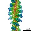


 Sample components
 Processing
 Electron microscopy
 FIELD EMISSION GUN