 Movie
Movie Controller

[English] 日本語
 Yorodumi
- EMDB-1450: Human T-lymphotropic virus-1 visualized at the virological synaps... -
+ Open data

- Basic information
| Entry | Database: EMDB / ID: EMD-1450 | |||||||||
|---|---|---|---|---|---|---|---|---|---|---|
| Title | Human T-lymphotropic virus-1 visualized at the virological synapse by electron tomography. | |||||||||
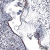
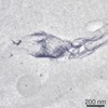
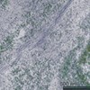
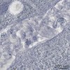
 Map data Map data | The tomogram shows a virological synapse between a naturally HTVL-1 infected CD4+ T lymphocyte (right) and a target cell (left). Two electron dense (anti-gag labeled) HTLV-1 particles can be seen in the synaptic cleft. | |||||||||
 Sample Sample |
| |||||||||
| Biological species |  Homo sapiens (human) Homo sapiens (human) | |||||||||
| Method | electron tomography / negative staining / Resolution: 50.0 Å | |||||||||
 Authors Authors | Majorovits E / Nejmeddine M / Tanaka Y / Taylor GP / Fuller SD / Bangham CRM | |||||||||
 Citation Citation | Journal: PLoS One / Year: 2008 Title: Human T-lymphotropic virus-1 visualized at the virological synapse by electron tomography. Authors: Endre Majorovits / Mohamed Nejmeddine / Yuetsu Tanaka / Graham P Taylor / Stephen D Fuller / Charles R M Bangham /  Abstract: Human T-lymphotropic virus 1 (HTLV-1) is transmitted directly between cells via an organized cell-cell contact called a virological synapse (VS). The VS has been studied by light microscopy, but the ...Human T-lymphotropic virus 1 (HTLV-1) is transmitted directly between cells via an organized cell-cell contact called a virological synapse (VS). The VS has been studied by light microscopy, but the ultrastructure of the VS and the nature of the transmitted viral particle have remained unknown. Cell-free enveloped virions of HTLV-1 are undetectable in the serum of individuals infected with the human T-lymphotropic virus 1 (HTLV-1) and during in vitro culture of naturally infected lymphocytes. However, the viral envelope protein is required for infectivity of HTLV-1, suggesting that complete, enveloped HTLV-1 virions are transferred across the synapse. Here, we use electron tomography combined with immunostaining of viral protein to demonstrate the presence of enveloped HTLV-1 particles within the VS formed between naturally infected lymphocytes. We show in 3D that HTLV-1 particles can be detected in multiple synaptic clefts at different locations simultaneously within the same VS. The synaptic clefts are surrounded by the tightly apposed plasma membranes of the two cells. HTLV-1 virions can contact the recipient cell membrane before detaching from the infected cell. The results show that the HTLV-1 virological synapse that forms spontaneously between lymphocytes of HTLV-1 infected individuals allows direct cell-cell transmission of the virus by triggered, directional release of enveloped HTLV-1 particles into confined intercellular spaces. | |||||||||
| History |
|
- Structure visualization
| Movie |
 Movie viewer Movie viewer |
|---|---|
| Structure viewer | EM map: SurfViewMolmilJmol/JSmol |
| Supplemental images |

- Downloads & links
-EMDB archive
| Map data | emd_1450.map.gz | 61 MB | EMDB map data format | |
|---|---|---|---|---|
| Header (meta data) | emd-1450-v30.xmlemd-1450.xml | 8.9 KB 8.9 KB | Display Display | EMDB header |
| Images |  1450.gif 1450.gif | 196.9 KB | ||
| Archive directory |  http://ftp.pdbj.org/pub/emdb/structures/EMD-1450ftp://ftp.pdbj.org/pub/emdb/structures/EMD-1450 http://ftp.pdbj.org/pub/emdb/structures/EMD-1450ftp://ftp.pdbj.org/pub/emdb/structures/EMD-1450 | HTTPS FTP |
-Validation report
| Summary document | emd_1450_validation.pdf.gz | 145.3 KB | Display | EMDB validaton report |
|---|---|---|---|---|
| Full document | emd_1450_full_validation.pdf.gz | 144.5 KB | Display | |
| Data in XML | emd_1450_validation.xml.gz | 4.7 KB | Display | |
| Arichive directory | https://ftp.pdbj.org/pub/emdb/validation_reports/EMD-1450ftp://ftp.pdbj.org/pub/emdb/validation_reports/EMD-1450 | HTTPS FTP |
-Related structure data
-Links
| EMDB pages | EMDB (EBI/PDBe) / EMDataResource |
|---|
-Map
| File | Download / File: emd_1450.map.gz / Format: CCP4 / Size: 156.2 MB / Type: IMAGE STORED AS FLOATING POINT NUMBER (4 BYTES) | ||||||||||||||||||||||||||||||||||||||||||||||||||||||||||||||||||||
|---|---|---|---|---|---|---|---|---|---|---|---|---|---|---|---|---|---|---|---|---|---|---|---|---|---|---|---|---|---|---|---|---|---|---|---|---|---|---|---|---|---|---|---|---|---|---|---|---|---|---|---|---|---|---|---|---|---|---|---|---|---|---|---|---|---|---|---|---|---|
| Annotation | The tomogram shows a virological synapse between a naturally HTVL-1 infected CD4+ T lymphocyte (right) and a target cell (left). Two electron dense (anti-gag labeled) HTLV-1 particles can be seen in the synaptic cleft. | ||||||||||||||||||||||||||||||||||||||||||||||||||||||||||||||||||||
| Voxel size | X: 33.6 Å / Y: 33.6 Å / Z: 25.9 Å | ||||||||||||||||||||||||||||||||||||||||||||||||||||||||||||||||||||
| Density |
| ||||||||||||||||||||||||||||||||||||||||||||||||||||||||||||||||||||
| Symmetry | Space group: 1 | ||||||||||||||||||||||||||||||||||||||||||||||||||||||||||||||||||||
| Details | EMDB XML:
CCP4 map header:
| ||||||||||||||||||||||||||||||||||||||||||||||||||||||||||||||||||||
-Supplemental data
- Sample components
-Entire : HTLV-1 virological snapse formed between CD4 T lymphocytes of HTL...
| Entire | Name: HTLV-1 virological snapse formed between CD4 T lymphocytes of HTLV-1 infeceted individual |
|---|---|
| Components |
|
-Supramolecule #1000: HTLV-1 virological snapse formed between CD4 T lymphocytes of HTL...
| Supramolecule | Name: HTLV-1 virological snapse formed between CD4 T lymphocytes of HTLV-1 infeceted individual type: sample / ID: 1000 / Number unique components: 1 |
|---|
-Supramolecule #1: virological synapse
| Supramolecule | Name: virological synapse / type: organelle_or_cellular_component / ID: 1 / Number of copies: 1 / Recombinant expression: No / Database: NCBI |
|---|---|
| Source (natural) | Organism: Homo sapiens (human) / synonym: human being / Cell: CD4 T lymphocyte / Organelle: Plasma membrane / Location in cell: Plasma membrane |
-Experimental details
-Structure determination
| Method | negative staining |
|---|---|
 Processing Processing | electron tomography |
-Sample preparation
| Buffer | pH: 7.2 / Details: sodium cacodylate buffer 0.1M |
|---|---|
| Staining | Type: NEGATIVE / Details: 1% OsO4 staining |
| Grid | Details: Formvar coated 2x1mm Cu slot grid |
| Vitrification | Cryogen name: NONE |
- Electron microscopy
| Microscope | FEI TECNAI F30 |
|---|---|
| Temperature | Average: 290 K |
| Specialist optics | Energy filter - Name: GIF / Energy filter - Lower energy threshold: 0.0 eV / Energy filter - Upper energy threshold: 25.0 eV |
| Date | Feb 26, 2007 |
| Image recording | Category: CCD / Film or detector model: GENERIC GATAN (2k x 2k) |
| Electron beam | Acceleration voltage: 300 kV / Electron source:  FIELD EMISSION GUN FIELD EMISSION GUN |
| Electron optics | Calibrated magnification: 10800 / Illumination mode: FLOOD BEAM / Imaging mode: BRIGHT FIELD / Cs: 2.2 mm / Nominal defocus max: 3.0 µm / Nominal defocus min: 3.0 µm / Nominal magnification: 9300 |
| Sample stage | Specimen holder: Gatan flip-flop holder / Specimen holder model: OTHER / Tilt series - Axis1 - Min angle: 65.0 ° / Tilt series - Axis1 - Max angle: 65 ° / Tilt series - Axis1 - Angle increment: 1 ° |
| Experimental equipment |  Model: Tecnai F30 / Image courtesy: FEI Company |
-Image processing
| Final reconstruction | Algorithm: OTHER / Resolution.type: BY AUTHOR / Resolution: 50.0 Å / Software - Name: IMOD / Number images used: 130 |
|---|
