- EMDB-1450: Human T-lymphotropic virus-1 visualized at the virological synaps... -
+
データを開く
IDまたはキーワード:
読み込み中...
-
基本情報
登録情報
データベース: EMDB / ID: EMD-1450
タイトル
Human T-lymphotropic virus-1 visualized at the virological synapse by electron tomography.
マップデータ
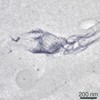
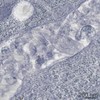
The tomogram shows a virological synapse between a naturally HTVL-1 infected CD4+ T lymphocyte (right) and a target cell (left). Two electron dense (anti-gag labeled) HTLV-1 particles can be seen in the synaptic cleft.
試料
試料: HTLV-1 virological snapse formed between CD4 T lymphocytes of HTLV-1 infeceted individual
ジャーナル: PLoS One / 年: 2008 タイトル: Human T-lymphotropic virus-1 visualized at the virological synapse by electron tomography. 著者: Endre Majorovits / Mohamed Nejmeddine / Yuetsu Tanaka / Graham P Taylor / Stephen D Fuller / Charles R M Bangham / 要旨: Human T-lymphotropic virus 1 (HTLV-1) is transmitted directly between cells via an organized cell-cell contact called a virological synapse (VS). The VS has been studied by light microscopy, but the ...Human T-lymphotropic virus 1 (HTLV-1) is transmitted directly between cells via an organized cell-cell contact called a virological synapse (VS). The VS has been studied by light microscopy, but the ultrastructure of the VS and the nature of the transmitted viral particle have remained unknown. Cell-free enveloped virions of HTLV-1 are undetectable in the serum of individuals infected with the human T-lymphotropic virus 1 (HTLV-1) and during in vitro culture of naturally infected lymphocytes. However, the viral envelope protein is required for infectivity of HTLV-1, suggesting that complete, enveloped HTLV-1 virions are transferred across the synapse. Here, we use electron tomography combined with immunostaining of viral protein to demonstrate the presence of enveloped HTLV-1 particles within the VS formed between naturally infected lymphocytes. We show in 3D that HTLV-1 particles can be detected in multiple synaptic clefts at different locations simultaneously within the same VS. The synaptic clefts are surrounded by the tightly apposed plasma membranes of the two cells. HTLV-1 virions can contact the recipient cell membrane before detaching from the infected cell. The results show that the HTLV-1 virological synapse that forms spontaneously between lymphocytes of HTLV-1 infected individuals allows direct cell-cell transmission of the virus by triggered, directional release of enveloped HTLV-1 particles into confined intercellular spaces.
ダウンロード / ファイル: emd_1450.map.gz / 形式: CCP4 / 大きさ: 156.2 MB / タイプ: IMAGE STORED AS FLOATING POINT NUMBER (4 BYTES)
注釈
The tomogram shows a virological synapse between a naturally HTVL-1 infected CD4+ T lymphocyte (right) and a target cell (left). Two electron dense (anti-gag labeled) HTLV-1 particles can be seen in the synaptic cleft.
ボクセルのサイズ
X: 33.6 Å / Y: 33.6 Å / Z: 25.9 Å
密度
最小 - 最大
-128.0 - 127.0
平均 (標準偏差)
17.9574 (±4.31085)
対称性
空間群: 1
詳細
EMDB XML:
マップ形状
Axis order
X
Y
Z
Origin
0
0
0
サイズ
1300
600
215
Spacing
1300
600
215
セル
A: 43680 Å / B: 20160 Å / C: 5568.5 Å α=β=γ: 90 °
CCP4マップ ヘッダ情報:
mode
envelope stored as signed bytes (from -128 lowest to 127 highest)
Å/pix. X/Y/Z
33.599996923077
33.6
25.9
M x/y/z
1300
600
215
origin x/y/z
0.000
0.000
0.000
length x/y/z
43679.996
20160.000
5568.500
α/β/γ
90.000
90.000
90.000
start NX/NY/NZ
-60
-60
-59
NX/NY/NZ
120
120
120
MAP C/R/S
1
2
3
start NC/NR/NS
0
0
0
NC/NR/NS
600
1300
215
D min/max/mean
-128.000
127.000
17.957
-
添付データ
-
試料の構成要素
-
全体 : HTLV-1 virological snapse formed between CD4 T lymphocytes of HTL...
全体
名称: HTLV-1 virological snapse formed between CD4 T lymphocytes of HTLV-1 infeceted individual
要素
試料: HTLV-1 virological snapse formed between CD4 T lymphocytes of HTLV-1 infeceted individual
細胞器官・細胞要素: virological synapse
-
超分子 #1000: HTLV-1 virological snapse formed between CD4 T lymphocytes of HTL...
超分子
名称: HTLV-1 virological snapse formed between CD4 T lymphocytes of HTLV-1 infeceted individual タイプ: sample / ID: 1000 / Number unique components: 1
 ムービー
ムービー コントローラー


 データを開く
 基本情報
基本情報 マップデータ
 試料
試料
 Homo sapiens (ヒト)
 データ登録者
 引用
引用
 構造の表示
構造の表示 ムービービューア
 ダウンロードとリンク
 1450.gif
 http://ftp.pdbj.org/pub/emdb/structures/EMD-1450
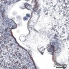
 試料の構成要素
 解析
解析 電子顕微鏡法
