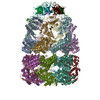
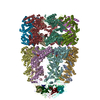
ジャーナル: Cell / 年: 2001 タイトル: ATP-bound states of GroEL captured by cryo-electron microscopy. 著者: N A Ranson / G W Farr / A M Roseman / B Gowen / W A Fenton / A L Horwich / H R Saibil / 要旨: The chaperonin GroEL drives its protein-folding cycle by cooperatively binding ATP to one of its two rings, priming that ring to become folding-active upon GroES binding, while simultaneously ...The chaperonin GroEL drives its protein-folding cycle by cooperatively binding ATP to one of its two rings, priming that ring to become folding-active upon GroES binding, while simultaneously discharging the previous folding chamber from the opposite ring. The GroEL-ATP structure, determined by cryo-EM and atomic structure fitting, shows that the intermediate domains rotate downward, switching their intersubunit salt bridge contacts from substrate binding to ATP binding domains. These observations, together with the effects of ATP binding to a GroEL-GroES-ADP complex, suggest structural models for the ATP-induced reduction in affinity for polypeptide and for cooperativity. The model for cooperativity, based on switching of intersubunit salt bridge interactions around the GroEL ring, may provide general insight into cooperativity in other ring complexes and molecular machines.
ダウンロード / ファイル: emd_1046.map.gz / 形式: CCP4 / 大きさ: 7.8 MB / タイプ: IMAGE STORED AS FLOATING POINT NUMBER (4 BYTES)
注釈
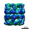
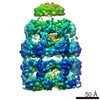
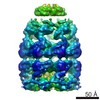
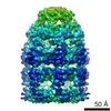
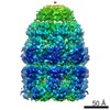
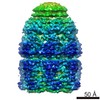
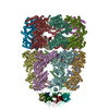
GroEL with GroES and ADP bound to one ring, and ATP bound to the other ring
ボクセルのサイズ
X=Y=Z: 2.8 Å
密度
表面レベル
登録者による: 0.029 / ムービー #1: 0.08
最小 - 最大
-0.04720771 - 0.27609465
平均 (標準偏差)
0.00606513 (±0.02973022)
対称性
空間群: 1
詳細
EMDB XML:
マップ形状
Axis order
X
Y
Z
Origin
0
0
0
サイズ
128
128
128
Spacing
128
128
128
セル
A=B=C: 358.4 Å α=β=γ: 90.0 °
CCP4マップ ヘッダ情報:
mode
Image stored as Reals
Å/pix. X/Y/Z
2.8
2.8
2.8
M x/y/z
128
128
128
origin x/y/z
0.000
0.000
0.000
length x/y/z
358.400
358.400
358.400
α/β/γ
90.000
90.000
90.000
start NX/NY/NZ
0
0
52
NX/NY/NZ
128
128
55
MAP C/R/S
1
2
3
start NC/NR/NS
0
0
0
NC/NR/NS
128
128
128
D min/max/mean
-0.047
0.276
0.006
-
添付データ
-
試料の構成要素
-
全体 : GroES-ADP7-GroEL-ATP7 from E.coli
全体
名称: GroES-ADP7-GroEL-ATP7 from E.coli
要素
試料: GroES-ADP7-GroEL-ATP7 from E.coli
タンパク質・ペプチド: GroEL
タンパク質・ペプチド: GroES
-
超分子 #1000: GroES-ADP7-GroEL-ATP7 from E.coli
超分子
名称: GroES-ADP7-GroEL-ATP7 from E.coli / タイプ: sample / ID: 1000 詳細: The complexes were prepared by pre-forming a GroEL-GroES-ADP complex, with 100 uM ADP, then adding an excess of single ring GroEL (SR1) to trap any released GroES; then 1 mM ATP was added. ...詳細: The complexes were prepared by pre-forming a GroEL-GroES-ADP complex, with 100 uM ADP, then adding an excess of single ring GroEL (SR1) to trap any released GroES; then 1 mM ATP was added. Any GroEL-GroES complexes remaining have ADP in the GroES-bound ring and ATP in the other ring, modelling the in vivo ATP-binding reaction. 集合状態: 14-mer / Number unique components: 2
pH: 7.5 詳細: 12.5 mM HEPES, 5 mM KCl, 5 mM MgCl2, 100 uM ADP, then 2x molar excess of single ring mutant of GroEL (SR1), then 1 mM ATP just before vitrification. See sample details for an explanation of ...詳細: 12.5 mM HEPES, 5 mM KCl, 5 mM MgCl2, 100 uM ADP, then 2x molar excess of single ring mutant of GroEL (SR1), then 1 mM ATP just before vitrification. See sample details for an explanation of the experimental design.
グリッド
詳細: holey carbon film
凍結
凍結剤: ETHANE / チャンバー内温度: 100 K / 装置: HOMEMADE PLUNGER / 詳細: Vitrification instrument: self made Timed resolved state: The sample was vitrified within a few seconds of manually mixing in ATP. 手法: Blot for 1 second before plunging
Protocol: Rigid body. The only movement relative to the crystal structure of GroEL-ES-ADP seen at this resolution is a twist of the free apical domains.
精密化
空間: REAL / プロトコル: RIGID BODY FIT
得られたモデル
PDB-1gru: SOLUTION STRUCTURE OF GROES-ADP7-GROEL-ATP7 COMPLEX BY CRYO-EM
 ムービー
ムービー コントローラー


 データを開く
 基本情報
基本情報 マップデータ
 試料
試料 機能・相同性情報
 データ登録者
 引用
引用
 構造の表示
構造の表示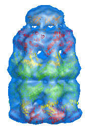
 ダウンロードとリンク
 1046.gif
 http://ftp.pdbj.org/pub/emdb/structures/EMD-1046
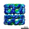


 試料の構成要素
 解析
解析 電子顕微鏡法
 FIELD EMISSION GUN