 Movie
Movie Controller

[English] 日本語
 Yorodumi
- EMDB-1020: Structure of a viral DNA gatekeeper at 10 A resolution by cryo-el... -
+ Open data

- Basic information
| Entry | Database: EMDB / ID: EMD-1020 | |||||||||
|---|---|---|---|---|---|---|---|---|---|---|
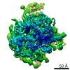
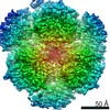
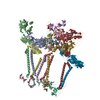
| Title | Structure of a viral DNA gatekeeper at 10 A resolution by cryo-electron microscopy. | |||||||||
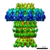
 Map data Map data | Viral DNA gatekeeper. Centre of symmetry : 50.0, 50.0, 50.0 | |||||||||
 Sample Sample |
| |||||||||
| Biological species |  Bacillus phage SPP1 (virus) Bacillus phage SPP1 (virus) | |||||||||
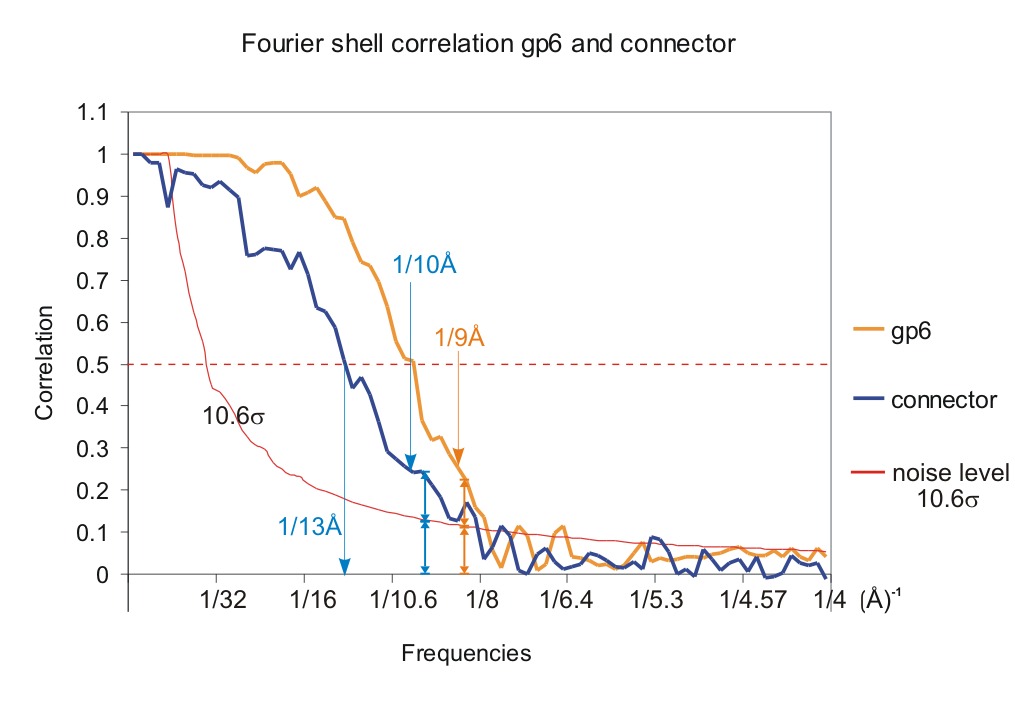
| Method | single particle reconstruction / cryo EM / Resolution: 10.0 Å | |||||||||
 Authors Authors | Orlova EV / Gowen B / Droege A / Stiege A / Weise F / Lurz R / van Heel M / Tavares P | |||||||||
 Citation Citation | Journal: EMBO J / Year: 2003 Title: Structure of a viral DNA gatekeeper at 10 A resolution by cryo-electron microscopy. Authors: Elena V Orlova / Brent Gowen / Anja Dröge / Asita Stiege / Frank Weise / Rudi Lurz / Marin van Heel / Paulo Tavares /  Abstract: In tailed bacteriophages and herpes viruses, the viral DNA is packaged through the portal protein channel. Channel closure is essential to prevent DNA release after packaging. Here we present the ...In tailed bacteriophages and herpes viruses, the viral DNA is packaged through the portal protein channel. Channel closure is essential to prevent DNA release after packaging. Here we present the connector structure from bacteriophage SPP1 using cryo-electron microscopy and single particle analysis. The multiprotein complex comprises the portal protein gp6 and the head completion proteins gp15 and gp16. Although we show that gp6 in the connector has a fold similar to that of the isolated portal protein, we observe conformational changes in the region of gp6 exposed to the DNA-packaging ATPase and to gp15. This reorganization does not cause closure of the channel. The connector channel traverses the full height of gp6 and gp15, but it is closed by gp16 at the bottom of the complex. Gp16 acts as a valve whose closure prevents DNA leakage, while its opening is required for DNA release upon interaction of the virus with its host. | |||||||||
| History |
|
- Structure visualization
| Movie |
 Movie viewer Movie viewer |
|---|---|
| Structure viewer | EM map: SurfViewMolmilJmol/JSmol |
| Supplemental images |


- Downloads & links
-EMDB archive
| Map data | emd_1020.map.gz | 1.3 MB | EMDB map data format | |
|---|---|---|---|---|
| Header (meta data) | emd-1020-v30.xmlemd-1020.xml | 9.5 KB 9.5 KB | Display Display | EMDB header |
| Images |  1020.gifemd_1020.tifemd_emd_1020.tif 1020.gifemd_1020.tifemd_emd_1020.tif | 23.5 KB 588.8 KB 588.8 KB | ||
| Others | Resolution_Portal_Connector.tif | 143.8 KB | ||
| Archive directory |  http://ftp.pdbj.org/pub/emdb/structures/EMD-1020ftp://ftp.pdbj.org/pub/emdb/structures/EMD-1020 http://ftp.pdbj.org/pub/emdb/structures/EMD-1020ftp://ftp.pdbj.org/pub/emdb/structures/EMD-1020 | HTTPS FTP |
-Validation report
| Summary document | emd_1020_validation.pdf.gz | 235 KB | Display | EMDB validaton report |
|---|---|---|---|---|
| Full document | emd_1020_full_validation.pdf.gz | 234.1 KB | Display | |
| Data in XML | emd_1020_validation.xml.gz | 5.3 KB | Display | |
| Arichive directory | https://ftp.pdbj.org/pub/emdb/validation_reports/EMD-1020ftp://ftp.pdbj.org/pub/emdb/validation_reports/EMD-1020 | HTTPS FTP |
-Related structure data
-Links
| EMDB pages | EMDB (EBI/PDBe) / EMDataResource |
|---|
-Map
| File | Download / File: emd_1020.map.gz / Format: CCP4 / Size: 3.7 MB / Type: IMAGE STORED AS FLOATING POINT NUMBER (4 BYTES) | ||||||||||||||||||||||||||||||||||||||||||||||||||||||||||||
|---|---|---|---|---|---|---|---|---|---|---|---|---|---|---|---|---|---|---|---|---|---|---|---|---|---|---|---|---|---|---|---|---|---|---|---|---|---|---|---|---|---|---|---|---|---|---|---|---|---|---|---|---|---|---|---|---|---|---|---|---|---|
| Annotation | Viral DNA gatekeeper. Centre of symmetry : 50.0, 50.0, 50.0 | ||||||||||||||||||||||||||||||||||||||||||||||||||||||||||||
| Voxel size | X=Y=Z: 2.1 Å | ||||||||||||||||||||||||||||||||||||||||||||||||||||||||||||
| Density |
| ||||||||||||||||||||||||||||||||||||||||||||||||||||||||||||
| Symmetry | Space group: 1 | ||||||||||||||||||||||||||||||||||||||||||||||||||||||||||||
| Details | EMDB XML:
CCP4 map header:
| ||||||||||||||||||||||||||||||||||||||||||||||||||||||||||||
-Supplemental data
-Others
- Sample components
-Entire : Bacteriophage SPP1 portal protein gp6
| Entire | Name: Bacteriophage SPP1 portal protein gp6 |
|---|---|
| Components |
|
-Supramolecule #1000: Bacteriophage SPP1 portal protein gp6
| Supramolecule | Name: Bacteriophage SPP1 portal protein gp6 / type: sample / ID: 1000 Oligomeric state: isolated portal protein has 13- fold cyclical symmetry Number unique components: 1 |
|---|---|
| Molecular weight | Experimental: 57.3 KDa / Theoretical: 57.3 KDa |
-Macromolecule #1: Bacteriophage SPP1 portal protein gp6
| Macromolecule | Name: Bacteriophage SPP1 portal protein gp6 / type: protein_or_peptide / ID: 1 / Name.synonym: naive gp6 / Number of copies: 13 / Oligomeric state: C13 / Recombinant expression: Yes |
|---|---|
| Source (natural) | Organism: Bacillus phage SPP1 (virus) / synonym: Bacteriophage SPP1 |
| Recombinant expression | Organism:  |
-Experimental details
-Structure determination
| Method | cryo EM |
|---|---|
 Processing Processing | single particle reconstruction |
| Aggregation state | particle |
-Sample preparation
| Concentration | 1.5 mg/mL |
|---|---|
| Buffer | pH: 7.5 |
| Vitrification | Cryogen name: ETHANE / Instrument: HOMEMADE PLUNGER / Details: Vitrification instrument: Standard plunger / Method: Blot about 2-3 seconds before plunging. |
- Electron microscopy
| Microscope | FEI/PHILIPS CM200FEG/ST |
|---|---|
| Date | Jan 1, 1999 |
| Image recording | Category: FILM / Film or detector model: KODAK SO-163 FILM / Digitization - Scanner: PATCHWORK DENSITOMETER / Digitization - Sampling interval: 10.2 µm / Number real images: 15 / Average electron dose: 10 e/Å2 Details: Images were digitised using a patch work densitometer. Od range: 1.6 / Bits/pixel: 8 |
| Electron beam | Acceleration voltage: 200 kV / Electron source:  FIELD EMISSION GUN FIELD EMISSION GUN |
| Electron optics | Calibrated magnification: 48600 / Illumination mode: FLOOD BEAM / Imaging mode: BRIGHT FIELD / Cs: 2.1 mm / Nominal defocus max: 3.0 µm / Nominal defocus min: 1.7 µm / Nominal magnification: 50000 |
| Sample stage | Specimen holder: side-entry / Specimen holder model: GATAN LIQUID NITROGEN |
-Image processing
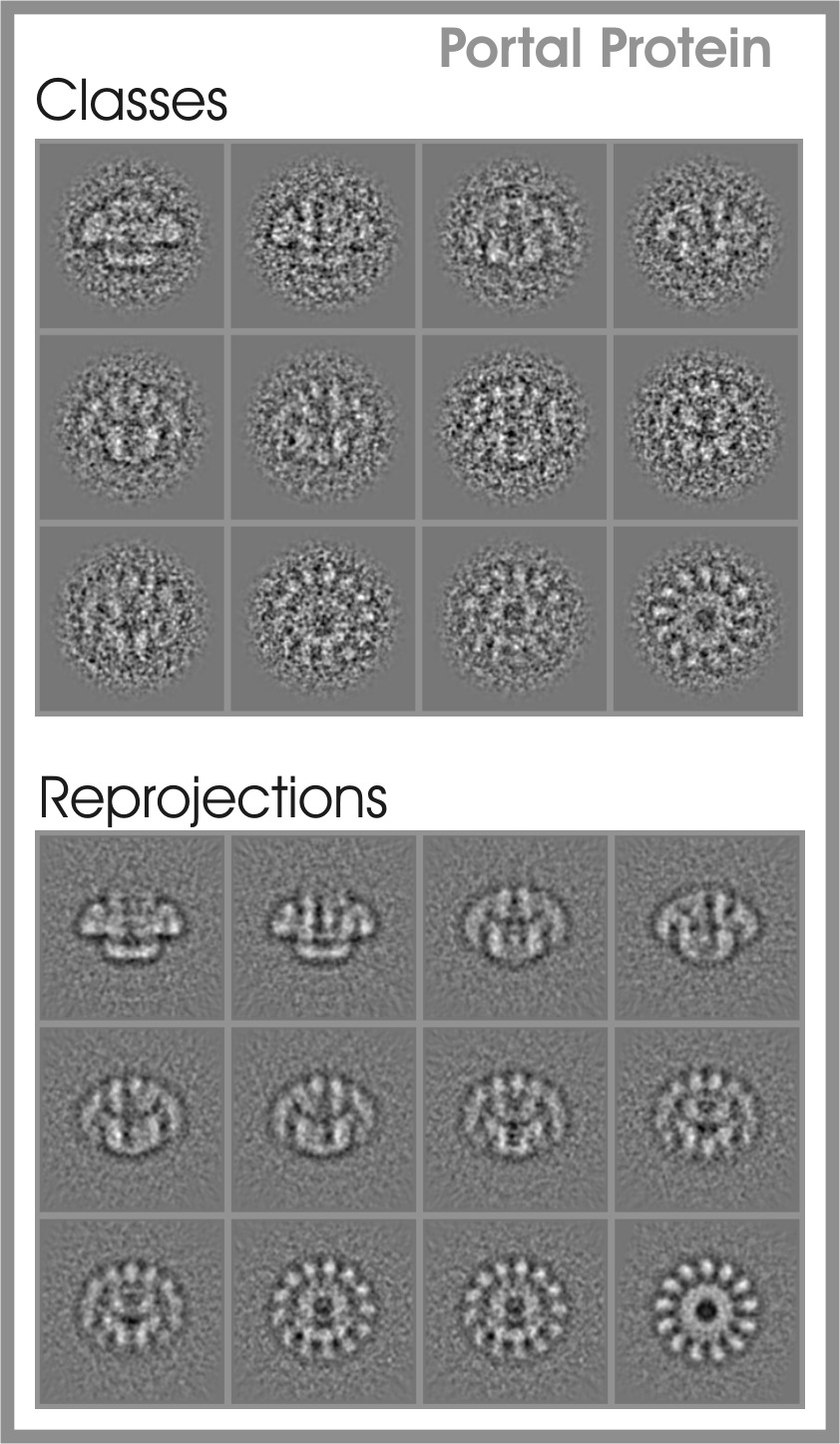
| Details | The number of classes analysed was 650. In the final reconstruction were used only 430. |
|---|---|
| CTF correction | Details: CTF correction of each particle |
| Final reconstruction | Applied symmetry - Point group: C13 (13 fold cyclic) / Algorithm: OTHER / Resolution.type: BY AUTHOR / Resolution: 10.0 Å / Resolution method: OTHER / Software - Name: IMAGIC / Number images used: 8000 |
| Final two d classification | Number classes: 430 |
-Atomic model buiding 1
| Software | Name: Molrep |
|---|---|
| Details | Protocol: Angular reconstitution |
