Movie
Movie Controller

[English] 日本語
 Yorodumi
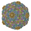
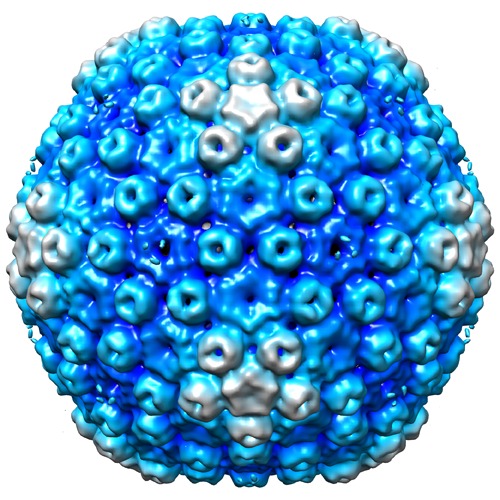
- EMDB-6043: Structure of the Head of Bacteriophage Basilisk to 18 Angstrom re... -
+ Open data

- Basic information
| Entry | Database: EMDB / ID: EMD-6043 | |||||||||
|---|---|---|---|---|---|---|---|---|---|---|
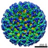
| Title | Structure of the Head of Bacteriophage Basilisk to 18 Angstrom resolution | |||||||||
 Map data Map data | Reconstruction of Basilisk Bacteriophage Head | |||||||||
 Sample Sample |
| |||||||||
 Keywords Keywords | bacteriophage / icosahedron / triangulation number / T=9 / capsid / siphovirus / HK97 fold / Bacillus cereus | |||||||||
| Biological species |  Bacillus phage Basilisk (virus) Bacillus phage Basilisk (virus) | |||||||||
| Method | single particle reconstruction / cryo EM / negative staining / Resolution: 18.0 Å | |||||||||
 Authors Authors | Grose JH / Belnap DM / Jensen JD / Mathis AD / Prince JT / Merrill B / Burnett SH / Breakwell DP | |||||||||
 Citation Citation | Journal: J Virol / Year: 2014 Title: The genomes, proteomes, and structures of three novel phages that infect the Bacillus cereus group and carry putative virulence factors. Authors: Julianne H Grose / David M Belnap / Jordan D Jensen / Andrew D Mathis / John T Prince / Bryan D Merrill / Sandra H Burnett / Donald P Breakwell /  Abstract: This article reports the results of studying three novel bacteriophages, JL, Shanette, and Basilisk, which infect the pathogen Bacillus cereus and carry genes that may contribute to its pathogenesis. ...This article reports the results of studying three novel bacteriophages, JL, Shanette, and Basilisk, which infect the pathogen Bacillus cereus and carry genes that may contribute to its pathogenesis. We analyzed host range and superinfection ability, mapped their genomes, and characterized phage structure by mass spectrometry and transmission electron microscopy (TEM). The JL and Shanette genomes were 96% similar and contained 217 open reading frames (ORFs) and 220 ORFs, respectively, while Basilisk has an unrelated genome containing 138 ORFs. Mass spectrometry revealed 23 phage particle proteins for JL and 15 for Basilisk, while only 11 and 4, respectively, were predicted to be present by sequence analysis. Structural protein homology to well-characterized phages suggested that JL and Shanette were members of the family Myoviridae, which was confirmed by TEM. The third phage, Basilisk, was similar only to uncharacterized phages and is an unrelated siphovirus. Cryogenic electron microscopy of this novel phage revealed a T=9 icosahedral capsid structure with the major capsid protein (MCP) likely having the same fold as bacteriophage HK97 MCP despite the lack of sequence similarity. Several putative virulence factors were encoded by these phage genomes, including TerC and TerD involved in tellurium resistance. Host range analysis of all three phages supports genetic transfer of such factors within the B. cereus group, including B. cereus, B. anthracis, and B. thuringiensis. This study provides a basis for understanding these three phages and other related phages as well as their contributions to the pathogenicity of B. cereus group bacteria. Importance: The Bacillus cereus group of bacteria contains several human and plant pathogens, including B. cereus, B. anthracis, and B. thuringiensis. Phages are intimately linked to the evolution of their bacterial hosts and often provide virulence factors, making the study of B. cereus phages important to understanding the evolution of pathogenic strains. Herein we provide the results of detailed study of three novel B. cereus phages, two highly related myoviruses (JL and Shanette) and an unrelated siphovirus (Basilisk). The detailed characterization of host range and superinfection, together with results of genomic, proteomic, and structural analyses, reveal several putative virulence factors as well as the ability of these phages to infect different pathogenic species. | |||||||||
| History |
|
- Structure visualization
| Movie |
 Movie viewer Movie viewer |
|---|---|
| Structure viewer | EM map: SurfViewMolmilJmol/JSmol |
| Supplemental images |
- Downloads & links
-EMDB archive
| Map data | emd_6043.map.gz | 69.2 MB | EMDB map data format | |
|---|---|---|---|---|
| Header (meta data) | emd-6043-v30.xmlemd-6043.xml | 10.4 KB 10.4 KB | Display Display | EMDB header |
| Images | emd_6043.tif | 767.1 KB | ||
| Archive directory |  http://ftp.pdbj.org/pub/emdb/structures/EMD-6043ftp://ftp.pdbj.org/pub/emdb/structures/EMD-6043 http://ftp.pdbj.org/pub/emdb/structures/EMD-6043ftp://ftp.pdbj.org/pub/emdb/structures/EMD-6043 | HTTPS FTP |
-Validation report
| Summary document | emd_6043_validation.pdf.gz | 78.3 KB | Display | EMDB validaton report |
|---|---|---|---|---|
| Full document | emd_6043_full_validation.pdf.gz | 77.3 KB | Display | |
| Data in XML | emd_6043_validation.xml.gz | 494 B | Display | |
| Arichive directory | https://ftp.pdbj.org/pub/emdb/validation_reports/EMD-6043ftp://ftp.pdbj.org/pub/emdb/validation_reports/EMD-6043 | HTTPS FTP |
-Related structure data
| Similar structure data |
|---|

-Links
| EMDB pages | EMDB (EBI/PDBe) / EMDataResource |
|---|
-Map
| File | Download / File: emd_6043.map.gz / Format: CCP4 / Size: 137 MB / Type: IMAGE STORED AS SIGNED INTEGER (2 BYTES) | ||||||||||||||||||||||||||||||||||||||||||||||||||||||||||||
|---|---|---|---|---|---|---|---|---|---|---|---|---|---|---|---|---|---|---|---|---|---|---|---|---|---|---|---|---|---|---|---|---|---|---|---|---|---|---|---|---|---|---|---|---|---|---|---|---|---|---|---|---|---|---|---|---|---|---|---|---|---|
| Annotation | Reconstruction of Basilisk Bacteriophage Head | ||||||||||||||||||||||||||||||||||||||||||||||||||||||||||||
| Voxel size | X=Y=Z: 2.02 Å | ||||||||||||||||||||||||||||||||||||||||||||||||||||||||||||
| Density |
| ||||||||||||||||||||||||||||||||||||||||||||||||||||||||||||
| Symmetry | Space group: 1 | ||||||||||||||||||||||||||||||||||||||||||||||||||||||||||||
| Details | EMDB XML:
CCP4 map header:
| ||||||||||||||||||||||||||||||||||||||||||||||||||||||||||||
-Supplemental data
- Sample components
-Entire : Bacteriophage Basilisk Head
| Entire | Name: Bacteriophage Basilisk Head |
|---|---|
| Components |
|
-Supramolecule #1000: Bacteriophage Basilisk Head
| Supramolecule | Name: Bacteriophage Basilisk Head / type: sample / ID: 1000 / Oligomeric state: icosahedral / Number unique components: 1 |
|---|
-Supramolecule #1: Bacillus phage Basilisk
| Supramolecule | Name: Bacillus phage Basilisk / type: virus / ID: 1 / Name.synonym: Bacteriophage Basilisk, Basilisk Phage Details: Bacteriophage Basilisk head component (NCBI taxonomy ID 1296654) Sci species name: Bacillus phage Basilisk / Database: NCBI / Virus type: VIRION / Virus isolate: SPECIES / Virus enveloped: No / Virus empty: No / Syn species name: Bacteriophage Basilisk, Basilisk Phage |
|---|---|
| Host (natural) | Organism:  |
| Virus shell | Shell ID: 1 / Name: capsid / Diameter: 780 Å / T number (triangulation number): 9 |
-Experimental details
-Structure determination
| Method | negative staining, cryo EM |
|---|---|
 Processing Processing | single particle reconstruction |
| Aggregation state | particle |
-Sample preparation
| Buffer | Details: PBS (phosphate-buffered saline) |
|---|---|
| Staining | Type: NEGATIVE / Details: no stain used |
| Grid | Details: holey carbon film (randomly sized holes) on copper grid |
| Vitrification | Cryogen name: ETHANE / Instrument: FEI VITROBOT MARK II Details: Blotting chamber (and sample) was maintained at 4 degrees Celsius before plunging; sample was applied with cold pipette tips. Humidity was 81-89%. Method: blotted 1-2 seconds before plunging |
- Electron microscopy
| Microscope | FEI TECNAI F20 |
|---|---|
| Date | Feb 14, 2014 |
| Image recording | Category: FILM / Film or detector model: KODAK SO-163 FILM / Digitization - Scanner: NIKON SUPER COOLSCAN 9000 / Digitization - Sampling interval: 6.35 µm / Number real images: 20 / Average electron dose: 4.4 e/Å2 / Details: 16-bit scanned images truncated to 8-bit / Bits/pixel: 16 |
| Tilt angle min | 0 |
| Tilt angle max | 0 |
| Electron beam | Acceleration voltage: 200 kV / Electron source:  FIELD EMISSION GUN FIELD EMISSION GUN |
| Electron optics | Calibrated magnification: 31400 / Illumination mode: FLOOD BEAM / Imaging mode: BRIGHT FIELD / Cs: 2.0 mm / Nominal defocus max: 3.3 µm / Nominal defocus min: 0.0 µm / Nominal magnification: 29000 |
| Sample stage | Specimen holder: nitrogen cooled / Specimen holder model: GATAN LIQUID NITROGEN |
| Experimental equipment |  Model: Tecnai F20 / Image courtesy: FEI Company |
-Image processing
| Final reconstruction | Algorithm: OTHER / Resolution.type: BY AUTHOR / Resolution: 18.0 Å / Resolution method: OTHER / Software - Name: AUTO3DEM / Number images used: 2085 |
|---|
-Atomic model buiding 1
| Initial model | PDB ID: |
|---|---|
| Refinement | Space: REAL / Protocol: RIGID BODY FIT |