Movie
Movie Controller

[English] 日本語
 Yorodumi
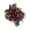
- PDB-5a6u: Native mammalian ribosome-bound Sec61 protein-conducting channel ... -
+ Open data

- Basic information
| Entry | Database: PDB / ID: 5a6u | |||||||||
|---|---|---|---|---|---|---|---|---|---|---|
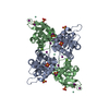
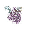
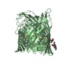
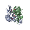
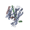
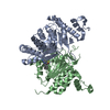
| Title | Native mammalian ribosome-bound Sec61 protein-conducting channel in the 'non-inserting' state | |||||||||
 Components Components |
| |||||||||
 Keywords Keywords | TRANSLATION / RIBOSOME / SEC61 / TRANSLOCON / ENDOPLASMIC RETICULUM / CRYOELECTRON TOMOGRAPHY / SUBTOMOGRAM ANALYSIS | |||||||||
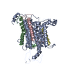
| Function / homology |  Function and homology information Function and homology informationInsertion of tail-anchored proteins into the endoplasmic reticulum membrane / membrane docking / endoplasmic reticulum Sec complex / pronephric nephron development / cotranslational protein targeting to membrane / Ssh1 translocon complex / Sec61 translocon complex / protein targeting to ER / protein-transporting ATPase activity / protein insertion into ER membrane ...Insertion of tail-anchored proteins into the endoplasmic reticulum membrane / membrane docking / endoplasmic reticulum Sec complex / pronephric nephron development / cotranslational protein targeting to membrane / Ssh1 translocon complex / Sec61 translocon complex / protein targeting to ER / protein-transporting ATPase activity / protein insertion into ER membrane / SRP-dependent cotranslational protein targeting to membrane, translocation / signal sequence binding / post-translational protein targeting to membrane, translocation / protein transmembrane transporter activity / guanyl-nucleotide exchange factor activity / phospholipid binding / ribosome binding / endoplasmic reticulum membrane / endoplasmic reticulum Similarity search - Function | |||||||||
| Biological species |  | |||||||||
| Method | ELECTRON MICROSCOPY / electron tomography / cryo EM / Resolution: 9 Å | |||||||||
 Authors Authors | Pfeffer, S. / Burbaum, L. / Unverdorben, P. / Pech, M. / Chen, Y. / Zimmermann, R. / Beckmann, R. / Foerster, F. | |||||||||
 Citation Citation | Journal: Nat Commun / Year: 2015 Title: Structure of the native Sec61 protein-conducting channel. Authors: Stefan Pfeffer / Laura Burbaum / Pia Unverdorben / Markus Pech / Yuxiang Chen / Richard Zimmermann / Roland Beckmann / Friedrich Förster /  Abstract: In mammalian cells, secretory and membrane proteins are translocated across or inserted into the endoplasmic reticulum (ER) membrane by the universally conserved protein-conducting channel Sec61, ...In mammalian cells, secretory and membrane proteins are translocated across or inserted into the endoplasmic reticulum (ER) membrane by the universally conserved protein-conducting channel Sec61, which has been structurally studied in isolated, detergent-solubilized states. Here we structurally and functionally characterize native, non-solubilized ribosome-Sec61 complexes on rough ER vesicles using cryo-electron tomography and ribosome profiling. Surprisingly, the 9-Å resolution subtomogram average reveals Sec61 in a laterally open conformation, even though the channel is not in the process of inserting membrane proteins into the lipid bilayer. In contrast to recent mechanistic models for polypeptide translocation and insertion, our results indicate that the laterally open conformation of Sec61 is the only conformation present in the ribosome-bound translocon complex, independent of its functional state. Consistent with earlier functional studies, our structure suggests that the ribosome alone, even without a nascent chain, is sufficient for lateral opening of Sec61 in a lipid environment. | |||||||||
| History |
|
- Structure visualization
| Movie |
Movie viewer |
|---|---|
| Structure viewer | Molecule: MolmilJmol/JSmol |
- Downloads & links
-Download
| PDBx/mmCIF format | 5a6u.cif.gz | 172.9 KB | Display | PDBx/mmCIF format |
|---|---|---|---|---|
| PDB format | pdb5a6u.ent.gz | 134.9 KB | Display | PDB format |
| PDBx/mmJSON format | 5a6u.json.gz | Tree view | PDBx/mmJSON format | |
| Others |  Other downloads Other downloads |
-Validation report
| Summary document | 5a6u_validation.pdf.gz | 688.1 KB | Display | wwPDB validaton report |
|---|---|---|---|---|
| Full document | 5a6u_full_validation.pdf.gz | 703.3 KB | Display | |
| Data in XML | 5a6u_validation.xml.gz | 22 KB | Display | |
| Data in CIF | 5a6u_validation.cif.gz | 31.6 KB | Display | |
| Arichive directory | https://data.pdbj.org/pub/pdb/validation_reports/a6/5a6uftp://data.pdbj.org/pub/pdb/validation_reports/a6/5a6u | HTTPS FTP |
-Related structure data
| Related structure data | 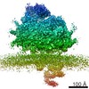 3068MC 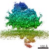 3069C 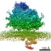 3070C 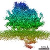 3071C 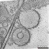 3072C M: map data used to model this data C: citing same article ( |
|---|---|
| Similar structure data |
-Links
 PDBj
PDBj

- Assembly
| Deposited unit | 
|
|---|---|
| 1 |
|
-Components
| #1: Protein | Mass: 49321.688 Da / Num. of mol.: 1 / Fragment: UNP RESIDUES 26-476 / Source method: isolated from a natural source / Source: (natural) |
|---|---|
| #2: Protein/peptide | Mass: 3992.791 Da / Num. of mol.: 1 / Fragment: UNP RESIDUES 61-96 / Source method: isolated from a natural source / Source: (natural) |
| #3: Protein | Mass: 7019.456 Da / Num. of mol.: 1 / Fragment: UNP RESIDUES 7-68 / Source method: isolated from a natural source / Source: (natural) |
-Experimental details
-Experiment
| Experiment | Method: ELECTRON MICROSCOPY |
|---|---|
| EM experiment | Aggregation state: PARTICLE / 3D reconstruction method: electron tomography |
- Sample preparation
| Component | Name: ER-DERIVED MICROSOMES / Type: ORGANELLE OR CELLULAR COMPONENT |
|---|---|
| Buffer solution | Name: 20MM HEPES, 50MM KCL, 2MM MGCL2 / pH: 7.6 / Details: 20MM HEPES, 50MM KCL, 2MM MGCL2 |
| Specimen | Conc.: 2 mg/ml / Embedding applied: NO / Shadowing applied: NO / Staining applied: NO / Vitrification applied: YES |
| Specimen support | Details: HOLEY CARBON |
| Vitrification | Instrument: FEI VITROBOT MARK IV / Cryogen name: ETHANE-PROPANE Details: VITRIFICATION 1 -- CRYOGEN- ETHANE-PROPANE MIXTURE, HUMIDITY- 70, INSTRUMENT- FEI VITROBOT MARK IV, METHOD- BLOT 3 SECONDS BEFORE PLUNGING., |
- Electron microscopy imaging
| Experimental equipment |  Model: Titan Krios / Image courtesy: FEI Company |
|---|---|
| Microscopy | Model: FEI TITAN KRIOS / Date: Jun 18, 2014 |
| Electron gun | Electron source:  FIELD EMISSION GUN / Accelerating voltage: 300 kV / Illumination mode: FLOOD BEAM FIELD EMISSION GUN / Accelerating voltage: 300 kV / Illumination mode: FLOOD BEAM |
| Electron lens | Mode: BRIGHT FIELD / Nominal defocus max: 4000 nm / Nominal defocus min: 3000 nm |
| Specimen holder | Tilt angle max: 20 ° / Tilt angle min: -20 ° |
| Image recording | Electron dose: 30 e/Å2 / Film or detector model: GATAN K2 (4k x 4k) |
| Image scans | Num. digital images: 2000 |
- Processing
| EM software |
| |||||||||||||||
|---|---|---|---|---|---|---|---|---|---|---|---|---|---|---|---|---|
| CTF correction | Details: EACH TILT IMAGE | |||||||||||||||
| Symmetry | Point symmetry: C1 (asymmetric) | |||||||||||||||
| 3D reconstruction | Resolution: 9 Å / Num. of particles: 17653 / Nominal pixel size: 2.62 Å / Actual pixel size: 2.62 Å Magnification calibration: CROSS- -CORRELATION WITH RIBOSOME DENSITIES Details: WEIGHTED BACKPROJECTION AND CONSTRAINED SUBTOMOGRAM AVERAGING SUBMISSION BASED ON EXPERIMENTAL DATA FROM EMDB EMD-3068. (DEPOSITION ID: 13544). Symmetry type: POINT | |||||||||||||||
| Atomic model building | Protocol: FLEXIBLE FIT / Space: REAL / Target criteria: PSEUDO-ENERGY / Details: METHOD--FLEXIBLE REFINEMENT PROTOCOL--CRYO-EM | |||||||||||||||
| Atomic model building | PDB-ID: 3J7Q Accession code: 3J7Q / Source name: PDB / Type: experimental model | |||||||||||||||
| Refinement | Highest resolution: 9 Å | |||||||||||||||
| Refinement step | Cycle: LAST / Highest resolution: 9 Å
|