 Movie
Movie Controller

[English] 日本語
 Yorodumi
- EMDB-5359: New mRNA-tRNA translocation intermediates revealed by cryoEM: Cla... -
+ Open data

- Basic information
| Entry | Database: EMDB / ID: EMD-5359 | |||||||||
|---|---|---|---|---|---|---|---|---|---|---|
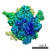
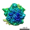
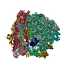
| Title | New mRNA-tRNA translocation intermediates revealed by cryoEM: Class 4A (partially rotated (I) 70S ribosome with A-site and P-site tRNAs) | |||||||||
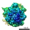
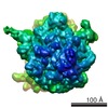
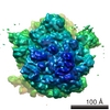
 Map data Map data | Cryo-revealed 70S ribosome partially rotated (i) (Class 4A) | |||||||||
 Sample Sample |
| |||||||||
 Keywords Keywords | ribosome / 70S / partially rotated / intermediates / translocation / tRNA | |||||||||
| Function / homology |  Function and homology information Function and homology informationnegative regulation of cytoplasmic translational initiation / stringent response / ornithine decarboxylase inhibitor activity / transcription antitermination factor activity, RNA binding / misfolded RNA binding / Group I intron splicing / RNA folding / transcriptional attenuation / endoribonuclease inhibitor activity / RNA-binding transcription regulator activity ...negative regulation of cytoplasmic translational initiation / stringent response / ornithine decarboxylase inhibitor activity / transcription antitermination factor activity, RNA binding / misfolded RNA binding / Group I intron splicing / RNA folding / transcriptional attenuation / endoribonuclease inhibitor activity / RNA-binding transcription regulator activity / positive regulation of ribosome biogenesis / negative regulation of cytoplasmic translation / four-way junction DNA binding / translational termination / DnaA-L2 complex / translation repressor activity / negative regulation of translational initiation / negative regulation of DNA-templated DNA replication initiation / regulation of mRNA stability / mRNA regulatory element binding translation repressor activity / ribosome assembly / positive regulation of RNA splicing / assembly of large subunit precursor of preribosome / transcription elongation factor complex / cytosolic ribosome assembly / regulation of DNA-templated transcription elongation / DNA endonuclease activity / ribosomal large subunit assembly / transcription antitermination / response to reactive oxygen species / translational initiation / regulation of cell growth / DNA-templated transcription termination / maintenance of translational fidelity / response to radiation / mRNA 5'-UTR binding / large ribosomal subunit / ribosome biogenesis / ribosome binding / regulation of translation / ribosomal small subunit biogenesis / ribosomal small subunit assembly / small ribosomal subunit / small ribosomal subunit rRNA binding / transferase activity / 5S rRNA binding / large ribosomal subunit rRNA binding / cytosolic small ribosomal subunit / cytosolic large ribosomal subunit / cytoplasmic translation / tRNA binding / molecular adaptor activity / rRNA binding / negative regulation of translation / ribosome / structural constituent of ribosome / translation / response to antibiotic / negative regulation of DNA-templated transcription / mRNA binding / DNA binding / RNA binding / zinc ion binding / membrane / cytoplasm / cytosol Similarity search - Function | |||||||||
| Biological species |  | |||||||||
| Method | single particle reconstruction / cryo EM / Resolution: 14.7 Å | |||||||||
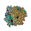
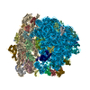
 Authors Authors | Agirrezabala X / Liao HY / Schreiner E / Fu J / Ortiz-Meoz RF / Schulten K / Green R / Frank J | |||||||||
 Citation Citation | Journal: Proc Natl Acad Sci U S A / Year: 2012 Title: Structural characterization of mRNA-tRNA translocation intermediates. Authors: Xabier Agirrezabala / Hstau Y Liao / Eduard Schreiner / Jie Fu / Rodrigo F Ortiz-Meoz / Klaus Schulten / Rachel Green / Joachim Frank /  Abstract: Cryo-EM analysis of a wild-type Escherichia coli pretranslocational sample has revealed the presence of previously unseen intermediate substates of the bacterial ribosome during the first phase of ...Cryo-EM analysis of a wild-type Escherichia coli pretranslocational sample has revealed the presence of previously unseen intermediate substates of the bacterial ribosome during the first phase of translocation, characterized by intermediate intersubunit rotations, L1 stalk positions, and tRNA configurations. Furthermore, we describe the domain rearrangements in quantitative terms, which has allowed us to characterize the processivity and coordination of the conformational reorganization of the ribosome, along with the associated changes in tRNA ribosome-binding configuration. The results are consistent with the view of the ribosome as a molecular machine employing Brownian motion to reach a functionally productive state via a series of substates with incremental changes in conformation. | |||||||||
| History |
|
- Structure visualization
| Movie |
Movie viewer |
|---|---|
| Structure viewer | EM map: SurfViewMolmilJmol/JSmol |
| Supplemental images |

- Downloads & links
-EMDB archive
| Map data | emd_5359.map.gz | 54.8 MB | EMDB map data format | |
|---|---|---|---|---|
| Header (meta data) | emd-5359-v30.xmlemd-5359.xml | 11.5 KB 11.5 KB | Display Display | EMDB header |
| Images | 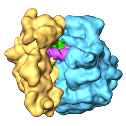 emd_5359_1.gif emd_5359_1.gif | 84.8 KB | ||
| Archive directory |  http://ftp.pdbj.org/pub/emdb/structures/EMD-5359ftp://ftp.pdbj.org/pub/emdb/structures/EMD-5359 http://ftp.pdbj.org/pub/emdb/structures/EMD-5359ftp://ftp.pdbj.org/pub/emdb/structures/EMD-5359 | HTTPS FTP |
-Validation report
| Summary document | emd_5359_validation.pdf.gz | 326.6 KB | Display | EMDB validaton report |
|---|---|---|---|---|
| Full document | emd_5359_full_validation.pdf.gz | 326.1 KB | Display | |
| Data in XML | emd_5359_validation.xml.gz | 5.9 KB | Display | |
| Arichive directory | https://ftp.pdbj.org/pub/emdb/validation_reports/EMD-5359ftp://ftp.pdbj.org/pub/emdb/validation_reports/EMD-5359 | HTTPS FTP |
-Related structure data
| Related structure data |  4v6oMC  5360C  5361C 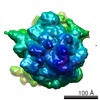 5362C 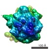 5363C 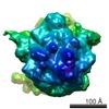 5364C 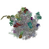 4v6nC 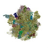 4v6pC 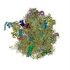 4v6qC 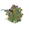 4v6rC 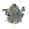 4v6sC M: atomic model generated by this map C: citing same article ( |
|---|---|
| Similar structure data |
-Links
| EMDB pages | EMDB (EBI/PDBe) / EMDataResource |
|---|---|
| Related items in Molecule of the Month |


-Map
| File | Download / File: emd_5359.map.gz / Format: CCP4 / Size: 58.2 MB / Type: IMAGE STORED AS FLOATING POINT NUMBER (4 BYTES) | ||||||||||||||||||||||||||||||||||||||||||||||||||||||||||||||||||||
|---|---|---|---|---|---|---|---|---|---|---|---|---|---|---|---|---|---|---|---|---|---|---|---|---|---|---|---|---|---|---|---|---|---|---|---|---|---|---|---|---|---|---|---|---|---|---|---|---|---|---|---|---|---|---|---|---|---|---|---|---|---|---|---|---|---|---|---|---|---|
| Annotation | Cryo-revealed 70S ribosome partially rotated (i) (Class 4A) | ||||||||||||||||||||||||||||||||||||||||||||||||||||||||||||||||||||
| Voxel size | X=Y=Z: 1.5 Å | ||||||||||||||||||||||||||||||||||||||||||||||||||||||||||||||||||||
| Density |
| ||||||||||||||||||||||||||||||||||||||||||||||||||||||||||||||||||||
| Symmetry | Space group: 1 | ||||||||||||||||||||||||||||||||||||||||||||||||||||||||||||||||||||
| Details | EMDB XML:
CCP4 map header:
| ||||||||||||||||||||||||||||||||||||||||||||||||||||||||||||||||||||
-Supplemental data
- Sample components
-Entire : Trp-tRNA-EFTu-GDP-kir-70S ribosome
| Entire | Name: Trp-tRNA-EFTu-GDP-kir-70S ribosome |
|---|---|
| Components |
|
-Supramolecule #1000: Trp-tRNA-EFTu-GDP-kir-70S ribosome
| Supramolecule | Name: Trp-tRNA-EFTu-GDP-kir-70S ribosome / type: sample / ID: 1000 / Oligomeric state: monomeric / Number unique components: 3 |
|---|---|
| Molecular weight | Experimental: 2.8 MDa / Theoretical: 2.8 MDa |
-Supramolecule #1: 70S ribosome
| Supramolecule | Name: 70S ribosome / type: complex / ID: 1 / Recombinant expression: No / Database: NCBI / Ribosome-details: ribosome-prokaryote: ALL |
|---|---|
| Source (natural) | Organism: |
-Experimental details
-Structure determination
| Method | cryo EM |
|---|---|
 Processing Processing | single particle reconstruction |
| Aggregation state | particle |
-Sample preparation
| Concentration | 0.03 mg/mL |
|---|---|
| Buffer | pH: 7.5 Details: HiFi buffer (50 mM Tris-HCl, pH 7.5, 70mM NH4Cl, 30 mM KCl, 3.5 mM MgCl2, 0.5 mM spermidine, 8mM putrescine, 2 mM DTT, 3.5 mM MgCl2) |
| Grid | Details: 200 mesh |
| Vitrification | Cryogen name: NITROGEN / Chamber humidity: 90 % / Chamber temperature: 80 K / Instrument: OTHER / Details: Vitrification instrument: Vitrobot / Method: blot for 3 seconds |
- Electron microscopy
| Microscope | FEI TECNAI F30 |
|---|---|
| Temperature | Min: 80.7 K / Max: 80.7 K / Average: 80.7 K |
| Alignment procedure | Legacy - Astigmatism: objective corrected at 100,000 times magnification Legacy - Electron beam tilt params: no tilt |
| Specialist optics | Energy filter - Name: no filter |
| Details | automated data collection system AutoEMation (CCD mag. 100000x) |
| Date | May 1, 2007 |
| Image recording | Category: CCD / Film or detector model: TVIPS TEMCAM-F415 (4k x 4k) / Average electron dose: 25 e/Å2 |
| Tilt angle min | 0 |
| Tilt angle max | 0 |
| Electron beam | Acceleration voltage: 300 kV / Electron source:  FIELD EMISSION GUN FIELD EMISSION GUN |
| Electron optics | Calibrated magnification: 58269 / Illumination mode: FLOOD BEAM / Imaging mode: BRIGHT FIELD / Cs: 2.26 mm / Nominal defocus max: 4.0 µm / Nominal defocus min: 1.2 µm / Nominal magnification: 59000 |
| Sample stage | Specimen holder: cartridge / Specimen holder model: OTHER |
| Experimental equipment |  Model: Tecnai F30 / Image courtesy: FEI Company |
-Image processing
| CTF correction | Details: defocus groups, Wiener filter |
|---|---|
| Final reconstruction | Resolution.type: BY AUTHOR / Resolution: 14.7 Å / Resolution method: FSC 0.5 CUT-OFF / Software - Name: XMIPP, SPIDER / Details: Classification using ML3D / Number images used: 21000 |
-Atomic model buiding 1
| Initial model | PDB ID:  2i2u |
|---|---|
| Software | Name: MDFF |
| Details | Protocol: Molecular Dynamics based flexible fitting |
| Refinement | Space: REAL / Protocol: FLEXIBLE FIT / Target criteria: RMSD, cross correlation |
| Output model | PDB-4v6o: |
-Atomic model buiding 2
| Initial model | PDB ID: 2i2v |
|---|---|
| Software | Name: MDFF |
| Details | Protocol: Molecular Dynamics based flexible fitting |
| Refinement | Space: REAL / Protocol: FLEXIBLE FIT / Target criteria: RMSD, cross correlation |
| Output model | PDB-4v6o: |
