 Movie
Movie Controller


+ Open data

- Basic information
| Entry | Database: EMDB / ID: EMD-5223 | |||||||||
|---|---|---|---|---|---|---|---|---|---|---|
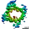
| Title | Human Ndc80 Bonsai Decorated Microtubule | |||||||||
 Map data Map data | Real space helical reconstruction of the human Ndc80 bonsai decorated microtubule | |||||||||
 Sample Sample |
| |||||||||
 Keywords Keywords | Ndc80 / HEC1 / NUF2 / tubulin / kinetochore / mitosis / calponin homology domain / microtubule | |||||||||
| Function / homology |  Function and homology information Function and homology informationG2/MI transition of meiotic cell cycle / kinetochore adaptor activity / skeletal muscle satellite cell proliferation / kinetochore => GO:0000776 / Ndc80 complex / kinetochore organization / metaphase chromosome alignment / positive regulation of mitotic cell cycle spindle assembly checkpoint / meiotic chromosome segregation / attachment of spindle microtubules to kinetochore ...G2/MI transition of meiotic cell cycle / kinetochore adaptor activity / skeletal muscle satellite cell proliferation / kinetochore => GO:0000776 / Ndc80 complex / kinetochore organization / metaphase chromosome alignment / positive regulation of mitotic cell cycle spindle assembly checkpoint / meiotic chromosome segregation / attachment of spindle microtubules to kinetochore / centrosome duplication / outer kinetochore / attachment of mitotic spindle microtubules to kinetochore / spindle assembly involved in female meiosis I / positive regulation of axon guidance / cell cycle / mitotic spindle assembly checkpoint signaling / establishment of mitotic spindle orientation / mitotic sister chromatid segregation / microtubule-based process / chromosome, centromeric region / cyclin binding / Amplification of signal from unattached kinetochores via a MAD2 inhibitory signal / Mitotic Prometaphase / EML4 and NUDC in mitotic spindle formation / Resolution of Sister Chromatid Cohesion / mitotic spindle organization / chromosome segregation / RHO GTPases Activate Formins / Hydrolases; Acting on acid anhydrides; Acting on GTP to facilitate cellular and subcellular movement / regulation of protein stability / structural constituent of cytoskeleton / kinetochore / microtubule cytoskeleton organization / Separation of Sister Chromatids / microtubule cytoskeleton / mitotic cell cycle / nervous system development / microtubule binding / microtubule / protein heterodimerization activity / cell division / GTPase activity / centrosome / protein-containing complex binding / nucleolus / GTP binding / nucleoplasm / identical protein binding / membrane / nucleus / metal ion binding / cytoplasm / cytosol Similarity search - Function | |||||||||
| Biological species |   Homo sapiens (human) Homo sapiens (human) | |||||||||
| Method | helical reconstruction / cryo EM / Resolution: 8.6 Å | |||||||||
 Authors Authors | Alushin GM / Ramey VH / Pasqualato S / Ball DA / Grigorieff N / Musacchio A / Nogales E | |||||||||
 Citation Citation | Journal: Nature / Year: 2010 Title: The Ndc80 kinetochore complex forms oligomeric arrays along microtubules. Authors: Gregory M Alushin / Vincent H Ramey / Sebastiano Pasqualato / David A Ball / Nikolaus Grigorieff / Andrea Musacchio / Eva Nogales /  Abstract: The Ndc80 complex is a key site of regulated kinetochore-microtubule attachment (a process required for cell division), but the molecular mechanism underlying its function remains unknown. Here we ...The Ndc80 complex is a key site of regulated kinetochore-microtubule attachment (a process required for cell division), but the molecular mechanism underlying its function remains unknown. Here we present a subnanometre-resolution cryo-electron microscopy reconstruction of the human Ndc80 complex bound to microtubules, sufficient for precise docking of crystal structures of the component proteins. We find that the Ndc80 complex binds the microtubule with a tubulin monomer repeat, recognizing α- and β-tubulin at both intra- and inter-tubulin dimer interfaces in a manner that is sensitive to tubulin conformation. Furthermore, Ndc80 complexes self-associate along protofilaments through interactions mediated by the amino-terminal tail of the NDC80 protein, which is the site of phospho-regulation by Aurora B kinase. The complex's mode of interaction with the microtubule and its oligomerization suggest a mechanism by which Aurora B could regulate the stability of load-bearing kinetochore-microtubule attachments. #1: Journal: CELL (CAMBRIDGE,MASS.) / Year: 2008Title: Implications for kinetochore-microtubule attachment from the structure of an engineered Ndc80 complex Authors: Ciferri C / Pasqualato S / Screpanti E / Varetti G / Santaguida S / Dos Reis G / Maiolica A / Polka J / De Luca JG / De Wulf P / Salek M / Rappsilber J / Moores CA / Salmon ED / Musacchio A #2: Journal: J.MOL.BIOL. / Year: 2001Title: Refined structure of alpha-beta tubulin from zinc-induced sheets stabilized with taxol Authors: Lowe J / Li H / Downing KH / Nogales E | |||||||||
| History |
|
- Structure visualization
| Movie |
Movie viewer |
|---|---|
| Structure viewer | EM map: SurfViewMolmilJmol/JSmol |
| Supplemental images |

- Downloads & links
-EMDB archive
| Map data | emd_5223.map.gz | 17.2 MB | EMDB map data format | |
|---|---|---|---|---|
| Header (meta data) | emd-5223-v30.xmlemd-5223.xml | 17.3 KB 17.3 KB | Display Display | EMDB header |
| Images | 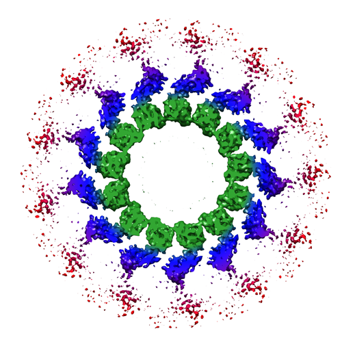 emd_5223_1.png emd_5223_1.png | 194.1 KB | ||
| Archive directory |  http://ftp.pdbj.org/pub/emdb/structures/EMD-5223ftp://ftp.pdbj.org/pub/emdb/structures/EMD-5223 http://ftp.pdbj.org/pub/emdb/structures/EMD-5223ftp://ftp.pdbj.org/pub/emdb/structures/EMD-5223 | HTTPS FTP |
-Validation report
| Summary document | emd_5223_validation.pdf.gz | 376.7 KB | Display | EMDB validaton report |
|---|---|---|---|---|
| Full document | emd_5223_full_validation.pdf.gz | 376.3 KB | Display | |
| Data in XML | emd_5223_validation.xml.gz | 4.7 KB | Display | |
| Arichive directory | https://ftp.pdbj.org/pub/emdb/validation_reports/EMD-5223ftp://ftp.pdbj.org/pub/emdb/validation_reports/EMD-5223 | HTTPS FTP |
-Related structure data
| Related structure data | 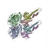 3iz0MC M: atomic model generated by this map C: citing same article ( |
|---|---|
| Similar structure data |


-Links
| EMDB pages | EMDB (EBI/PDBe) / EMDataResource |
|---|---|
| Related items in Molecule of the Month |


-Map
| File | Download / File: emd_5223.map.gz / Format: CCP4 / Size: 18 MB / Type: IMAGE STORED AS FLOATING POINT NUMBER (4 BYTES) | ||||||||||||||||||||||||||||||||||||||||||||||||||||||||||||||||||||
|---|---|---|---|---|---|---|---|---|---|---|---|---|---|---|---|---|---|---|---|---|---|---|---|---|---|---|---|---|---|---|---|---|---|---|---|---|---|---|---|---|---|---|---|---|---|---|---|---|---|---|---|---|---|---|---|---|---|---|---|---|---|---|---|---|---|---|---|---|---|
| Annotation | Real space helical reconstruction of the human Ndc80 bonsai decorated microtubule | ||||||||||||||||||||||||||||||||||||||||||||||||||||||||||||||||||||
| Voxel size | X=Y=Z: 2.48 Å | ||||||||||||||||||||||||||||||||||||||||||||||||||||||||||||||||||||
| Density |
| ||||||||||||||||||||||||||||||||||||||||||||||||||||||||||||||||||||
| Symmetry | Space group: 1 | ||||||||||||||||||||||||||||||||||||||||||||||||||||||||||||||||||||
| Details | EMDB XML:
CCP4 map header:
| ||||||||||||||||||||||||||||||||||||||||||||||||||||||||||||||||||||
-Supplemental data
- Sample components
-Entire : Human Ndc80 bonsai complex bound to the microtubule
| Entire | Name: Human Ndc80 bonsai complex bound to the microtubule |
|---|---|
| Components |
|
-Supramolecule #1000: Human Ndc80 bonsai complex bound to the microtubule
| Supramolecule | Name: Human Ndc80 bonsai complex bound to the microtubule / type: sample / ID: 1000 Details: Ndc80 bonsai is a heterodimer of Ndc80-Spc25 and Nuf2-Spc24 Tubulin is a heterodimer of alpha-beta tubulin Oligomeric state: 2 copies of the Ndc80 bonsai complex bind to each tubulin heterodimer Number unique components: 2 |
|---|---|
| Molecular weight | Theoretical: 260 KDa |
-Macromolecule #1: tubulin
| Macromolecule | Name: tubulin / type: protein_or_peptide / ID: 1 / Name.synonym: tubulin / Number of copies: 1 / Oligomeric state: heterodimer / Recombinant expression: No / Database: NCBI |
|---|---|
| Source (natural) | Organism: |
| Molecular weight | Theoretical: 110 KDa |
-Macromolecule #2: Ndc80-Spc25 Chimera
| Macromolecule | Name: Ndc80-Spc25 Chimera / type: protein_or_peptide / ID: 2 / Name.synonym: Ndc80-Spc25 Chimera Details: Chimera of Ndc80 residues 1-286 with Spc25 residues 118-224 Number of copies: 1 / Oligomeric state: heterodimer / Recombinant expression: Yes |
|---|---|
| Source (natural) | Organism: Homo sapiens (human) / synonym: human / Organelle: Nucleus / Location in cell: Kinetochore |
| Molecular weight | Theoretical: 45 KDa |
| Recombinant expression | Organism:  |
-Macromolecule #3: Nuf2-Spc24 Chimera
| Macromolecule | Name: Nuf2-Spc24 Chimera / type: protein_or_peptide / ID: 3 / Name.synonym: Nuf2-Spc24 Chimera Details: Chimera of Nuf2 residues 1-169 with Spc24 residues 122-197 Number of copies: 1 / Oligomeric state: heterodimer / Recombinant expression: Yes |
|---|---|
| Source (natural) | Organism: Homo sapiens (human) / synonym: Human / Organelle: Nucleus / Location in cell: Kinetochore |
| Molecular weight | Theoretical: 29 KDa |
| Recombinant expression | Organism: |
-Experimental details
-Structure determination
| Method | cryo EM |
|---|---|
 Processing Processing | helical reconstruction |
| Aggregation state | filament |
-Sample preparation
| Concentration | 0.25 mg/mL |
|---|---|
| Buffer | pH: 6.8 Details: 80mM PIPES, 1mM MgCl2, 1mM EGTA, 1mM DTT, 0.05% Nonidet P-40, 20uM taxol |
| Grid | Details: C-flat 1.2/1.3 |
| Vitrification | Cryogen name: ETHANE / Chamber humidity: 100 % / Instrument: OTHER / Details: Vitrification instrument: Vitrobot Method: 2ul of 0.25 mg per ml MTs applied to grid for 1 minute 4ul of 0.7 mg per ml Ndc80 bonsai added, 1 minute manually blotted, then another 4ul of Ndc80 applied 1 minute 2ul removed with pipetter ...Method: 2ul of 0.25 mg per ml MTs applied to grid for 1 minute 4ul of 0.7 mg per ml Ndc80 bonsai added, 1 minute manually blotted, then another 4ul of Ndc80 applied 1 minute 2ul removed with pipetter Blot for 2 seconds before plunging, 0mm offset |
- Electron microscopy
| Microscope | FEI TECNAI F20 |
|---|---|
| Alignment procedure | Legacy - Astigmatism: objective lens astigmatism corrected at 100Kx mag |
| Date | Mar 12, 2009 |
| Image recording | Category: FILM / Film or detector model: KODAK SO-163 FILM / Digitization - Scanner: NIKON SUPER COOLSCAN 9000 / Digitization - Sampling interval: 6.35 µm / Number real images: 100 / Average electron dose: 15 e/Å2 |
| Electron beam | Acceleration voltage: 200 kV / Electron source:  FIELD EMISSION GUN FIELD EMISSION GUN |
| Electron optics | Illumination mode: FLOOD BEAM / Imaging mode: BRIGHT FIELD / Cs: 2.2 mm / Nominal defocus max: 2.2 µm / Nominal defocus min: 1.2 µm / Nominal magnification: 50000 |
| Sample stage | Specimen holder: side-entry / Specimen holder model: GATAN LIQUID NITROGEN |
| Experimental equipment |  Model: Tecnai F20 / Image courtesy: FEI Company |
-Image processing
| Final reconstruction | Applied symmetry - Helical parameters - Δz: 9.54113 Å Applied symmetry - Helical parameters - Δ&Phi: 27.67984 ° Algorithm: OTHER / Resolution.type: BY AUTHOR / Resolution: 8.6 Å / Resolution method: FSC 0.143 CUT-OFF / Software - Name: Spider, FREALIGN Details: Particles were initially aligned using IHRSR protocol in SPIDER with naked MT as reference. Final reconstruction and CTF correction was performed with FREALIGN. A B-factor of -450 was ...Details: Particles were initially aligned using IHRSR protocol in SPIDER with naked MT as reference. Final reconstruction and CTF correction was performed with FREALIGN. A B-factor of -450 was applied with BFACTOR. FSC was calculated only for MT and Ndc80-NUF2 head, disordered outer head was excluded with soft mask. |
|---|
-Atomic model buiding 1
| Initial model | PDB ID: |
|---|---|
| Software | Name: Chimera |
| Details | Protocol: Rigid Body |
| Refinement | Space: REAL / Protocol: RIGID BODY FIT |
| Output model | PDB-3iz0: |

-Atomic model buiding 2
| Initial model | PDB ID: Chain - Chain ID: B |
|---|---|
| Software | Name: Chimera |
| Details | PDBEntryID_givenInChain. Protocol: Rigid Body. The pdb was manually edited to remove disordered residues in EM map it ends at residue four hundred thirty seven |
| Refinement | Space: REAL / Protocol: RIGID BODY FIT |
| Output model | PDB-3iz0: |

-Atomic model buiding 3
| Initial model | PDB ID: Chain - Chain ID: D |
|---|---|
| Software | Name: Chimera |
| Details | PDBEntryID_givenInChain. Protocol: Rigid Body. The pdb was manually edited to remove disordered residues in EM map it ends at residue one hundred fifty six |
| Refinement | Space: REAL / Protocol: RIGID BODY FIT |
| Output model | PDB-3iz0: |
