 ムービー
ムービー コントローラー


+ データを開く

- 基本情報
基本情報
| 登録情報 | データベース: EMDB / ID: EMD-5156 | |||||||||
|---|---|---|---|---|---|---|---|---|---|---|
| タイトル | Bovine Papillomavirus Type 1 (BPV1) cryo-EM reconstruction of L1 pentamer at 3.6A resolution after additional 6-fold NCS averaging within the icosahedral asymmetric unit. | |||||||||
 マップデータ マップデータ | This map is a result of additional 6-fold NCS averaging within the ASU. It shows the invariable part of the L1 pentamer and contains the core domain plus parts of the inter-pentamer C-terminal arm interface. | |||||||||
 試料 試料 |
| |||||||||
 キーワード キーワード | bovine papillomavirus BPV1 / major capsid protein L1 / single particle cryo-EM / high resolution / cervical cancer | |||||||||
| 機能・相同性 |  機能・相同性情報 機能・相同性情報T=7 icosahedral viral capsid / endocytosis involved in viral entry into host cell / host cell nucleus / virion attachment to host cell / structural molecule activity 類似検索 - 分子機能 | |||||||||
| 生物種 |  Bovine papillomavirus type 1 (パピローマウイルス) Bovine papillomavirus type 1 (パピローマウイルス) | |||||||||
| 手法 | 単粒子再構成法 / クライオ電子顕微鏡法 / 解像度: 3.6 Å | |||||||||
 データ登録者 データ登録者 | Wolf M / Garcea RL / Grigorieff N / Harrison SC | |||||||||
 引用 引用 | ジャーナル: Proc Natl Acad Sci U S A / 年: 2010 タイトル: Subunit interactions in bovine papillomavirus. 著者: Matthias Wolf / Robert L Garcea / Nikolaus Grigorieff / Stephen C Harrison /  要旨: Papillomaviruses, members of a group of dsDNA viruses associated with epithelial growths and tumors, have compact capsids assembled from 72 pentamers of the protein L1. We have determined the ...Papillomaviruses, members of a group of dsDNA viruses associated with epithelial growths and tumors, have compact capsids assembled from 72 pentamers of the protein L1. We have determined the structure of bovine papillomavirus by electron cryomicrosopy (cryoEM), at approximately 3.6 A resolution. The density map, obtained from single-particle analysis of approximately 4,000 particle images, shows the trace of the L1 polypeptide chain and reveals how the N- and C-terminal "arms" of a subunit (extensions from its beta-jelly-roll core) associate with a neighboring pentamer. Critical contacts come from the C-terminal arm, which loops out from the core of the subunit, forms contacts (including a disulfide) with two subunits in a neighboring pentamer, and reinserts into the pentamer from which it emanates. This trace corrects one feature of an earlier model. We discuss implications of the structure for virion assembly and for pathways of infectious viral entry. We suggest that it should be possible to obtain image reconstructions of comparable resolution from cryoEM images of asymmetric particles. From the work on papillomavirus described here, we estimate that such a reconstruction will require about 1.5 million images to achieve the same number of averaged asymmetric units; structural variability will increase this number substantially. | |||||||||
| 履歴 |
|
- 構造の表示
構造の表示
| ムービー |
ムービービューア |
|---|---|
| 構造ビューア | EMマップ: SurfViewMolmilJmol/JSmol |
| 添付画像 |

- ダウンロードとリンク
-EMDBアーカイブ
| マップデータ | emd_5156.map.gz | 1003.8 KB | EMDBマップデータ形式 | |
|---|---|---|---|---|
| ヘッダ (付随情報) | emd-5156-v30.xmlemd-5156.xml | 12.4 KB 12.4 KB | 表示 表示 | EMDBヘッダ |
| 画像 | 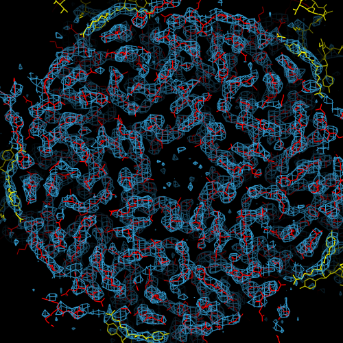 emd_5156_1.png emd_5156_1.png | 418.3 KB | ||
| アーカイブディレクトリ |  http://ftp.pdbj.org/pub/emdb/structures/EMD-5156ftp://ftp.pdbj.org/pub/emdb/structures/EMD-5156 http://ftp.pdbj.org/pub/emdb/structures/EMD-5156ftp://ftp.pdbj.org/pub/emdb/structures/EMD-5156 | HTTPS FTP |
-関連構造データ


-リンク
| EMDBのページ | EMDB (EBI/PDBe) / EMDataResource |
|---|---|
| 「今月の分子」の関連する項目 |


-マップ
| ファイル | ダウンロード / ファイル: emd_5156.map.gz / 形式: CCP4 / 大きさ: 6.4 MB / タイプ: IMAGE STORED AS FLOATING POINT NUMBER (4 BYTES) | ||||||||||||||||||||||||||||||||||||||||||||||||||||||||||||||||||||
|---|---|---|---|---|---|---|---|---|---|---|---|---|---|---|---|---|---|---|---|---|---|---|---|---|---|---|---|---|---|---|---|---|---|---|---|---|---|---|---|---|---|---|---|---|---|---|---|---|---|---|---|---|---|---|---|---|---|---|---|---|---|---|---|---|---|---|---|---|---|
| 注釈 | This map is a result of additional 6-fold NCS averaging within the ASU. It shows the invariable part of the L1 pentamer and contains the core domain plus parts of the inter-pentamer C-terminal arm interface. | ||||||||||||||||||||||||||||||||||||||||||||||||||||||||||||||||||||
| 投影像・断面図 | 画像のコントロール
画像は Spider により作成 これらの図は立方格子座標系で作成されたものです | ||||||||||||||||||||||||||||||||||||||||||||||||||||||||||||||||||||
| ボクセルのサイズ | X=Y=Z: 1.237 Å | ||||||||||||||||||||||||||||||||||||||||||||||||||||||||||||||||||||
| 密度 |
| ||||||||||||||||||||||||||||||||||||||||||||||||||||||||||||||||||||
| 対称性 | 空間群: 1 | ||||||||||||||||||||||||||||||||||||||||||||||||||||||||||||||||||||
| 詳細 | EMDB XML:
CCP4マップ ヘッダ情報:
| ||||||||||||||||||||||||||||||||||||||||||||||||||||||||||||||||||||
 Z (Sec.)
 Y (Row.)
 X (Col.)


-添付データ
- 試料の構成要素
-全体 : Bovine Papillomavirus Type 1 (BPV1), full virion
| 全体 | 名称: Bovine Papillomavirus Type 1 (BPV1), full virion |
|---|---|
| 要素 |
|
-超分子 #1000: Bovine Papillomavirus Type 1 (BPV1), full virion
| 超分子 | 名称: Bovine Papillomavirus Type 1 (BPV1), full virion / タイプ: sample / ID: 1000 / 詳細: theoretical MM for L1 only / 集合状態: T7D icosahedral assembly / Number unique components: 1 |
|---|---|
| 分子量 | 理論値: 19.4 MDa |
-超分子 #1: Bovine papillomavirus type 1
| 超分子 | 名称: Bovine papillomavirus type 1 / タイプ: virus / ID: 1 / Name.synonym: BPV1 / NCBI-ID: 10559 / 生物種: Bovine papillomavirus type 1 / データベース: NCBI / ウイルスタイプ: VIRION / ウイルス・単離状態: SEROTYPE / ウイルス・エンベロープ: No / ウイルス・中空状態: No / Syn species name: BPV1 |
|---|---|
| 宿主 | 生物種:  |
| 分子量 | 理論値: 19.4 MDa |
| ウイルス殻 | Shell ID: 1 / 名称: L1 / 直径: 600 Å / T番号(三角分割数): 7 |
-実験情報
-構造解析
| 手法 | クライオ電子顕微鏡法 |
|---|---|
 解析 解析 | 単粒子再構成法 |
| 試料の集合状態 | particle |
-試料調製
| 濃度 | 1.5 mg/mL |
|---|---|
| 緩衝液 | pH: 6.2 / 詳細: 20mM Tris pH6.2, 100mM NaCl, 0.5mM CaCl2 |
| グリッド | 詳細: quantifoil CF-1/2-4C |
| 凍結 | 凍結剤: ETHANE / チャンバー内湿度: 99 % / チャンバー内温度: 70 K / 装置: HOMEMADE PLUNGER 詳細: Vitrification instrument: manual plunger. vitrification carried out in cold room. 手法: blot 3.5ul at 4C for 25sec from carbon side with Whatman Nr.40 |
- 電子顕微鏡法
| 顕微鏡 | FEI TECNAI F30 |
|---|---|
| 温度 | 最低: 77 K / 最高: 77 K / 平均: 77 K |
| アライメント法 | Legacy - 非点収差: obj lens astig was corrected at 700,000x |
| 詳細 | obj aperture cutoff at 2.4A |
| 日付 | 2008年12月7日 |
| 撮影 | カテゴリ: FILM / フィルム・検出器のモデル: KODAK SO-163 FILM / デジタル化 - スキャナー: ZEISS SCAI / デジタル化 - サンプリング間隔: 7 µm / 実像数: 49 / 平均電子線量: 25 e/Å2 / 詳細: combination of 3 channels / Od range: 1.4 / ビット/ピクセル: 8 |
| 電子線 | 加速電圧: 300 kV / 電子線源:  FIELD EMISSION GUN FIELD EMISSION GUN |
| 電子光学系 | 倍率(補正後): 56588 / 照射モード: FLOOD BEAM / 撮影モード: BRIGHT FIELD / Cs: 2.0 mm / 最大 デフォーカス(公称値): 2.9 µm / 最小 デフォーカス(公称値): 1.8 µm / 倍率(公称値): 59000 |
| 試料ステージ | 試料ホルダー: side entry, eucentric / 試料ホルダーモデル: GATAN LIQUID NITROGEN |
| 実験機器 |  モデル: Tecnai F30 / 画像提供: FEI Company |
-画像解析
| CTF補正 | 詳細: ctfilt3 with individual particle adjustment |
|---|---|
| 最終 再構成 | アルゴリズム: OTHER / 解像度のタイプ: BY AUTHOR / 解像度: 3.6 Å / 解像度の算出法: FSC 0.143 CUT-OFF / ソフトウェア - 名称: frealign 詳細: This map was 6-fold (NCS)averaged within the icosahedral ASU. Ewald sphere correction. 使用した粒子像数: 3997 |
-原子モデル構築 1
| 初期モデル | PDB ID: |
|---|---|
| ソフトウェア | 名称: CNSsolve |
| 詳細 | Protocol: see 3D-fitting details. SigmaF and phase FOM were estimated from two halfset reconstructions (see publication). Refinement within the resolution range 15-3.5A. Isotropic B-factor correction, automatic bulk solvent parameter search, bulk-solvent correction, coordinate refinement by energy minimization, restrained individual B-factor refinement and unrestrained group B-factor refinement. Both icosahedaral and non-icosahedral capsid symmetry (NCS) were imposed, using icosahedral constraints and six-fold NCS restraints within the asymmetric unit defined in five NCS groups. We used default x-ray scattering factors and a maximum likelihood refinement target (MLHL) with amplitude and phase probability distribution. |
| 精密化 | 空間: RECIPROCAL / プロトコル: RIGID BODY FIT / 温度因子: 114 当てはまり具合の基準: MLHL target with ampl. and phase probability distribution |
| 得られたモデル |  PDB-3iyj: |

