 Movie
Movie Controller


+ Open data

- Basic information
| Entry | Database: PDB / ID: 3iyj | ||||||
|---|---|---|---|---|---|---|---|
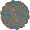
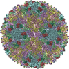
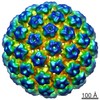
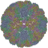
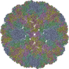
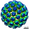
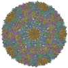
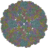
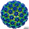
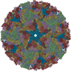
| Title | Bovine papillomavirus type 1 outer capsid | ||||||
 Components Components | Major capsid protein L1 | ||||||
 Keywords Keywords |  VIRUS / bovine papillomavirus BPV1 / major capsid protein L1 / single particle cryo-EM / high resolution / Capsid protein / Late protein / Virion VIRUS / bovine papillomavirus BPV1 / major capsid protein L1 / single particle cryo-EM / high resolution / Capsid protein / Late protein / Virion | ||||||
| Function / homology |  Function and homology information Function and homology informationT=7 icosahedral viral capsid / endocytosis involved in viral entry into host cell / host cell nucleus / structural molecule activity / virion attachment to host cell Similarity search - Function | ||||||
| Biological species |  Bovine papillomavirus type 1 Bovine papillomavirus type 1 | ||||||
| Method | ELECTRON MICROSCOPY / single particle reconstruction / cryo EM / Resolution: 4.2 Å | ||||||
 Authors Authors | Wolf, M. / Garcea, R.L. / Grigorieff, N. / Harrison, S.C. | ||||||
 Citation Citation | Journal: Proc Natl Acad Sci U S A / Year: 2010 Title: Subunit interactions in bovine papillomavirus. Authors: Matthias Wolf / Robert L Garcea / Nikolaus Grigorieff / Stephen C Harrison /  Abstract: Papillomaviruses, members of a group of dsDNA viruses associated with epithelial growths and tumors, have compact capsids assembled from 72 pentamers of the protein L1. We have determined the ...Papillomaviruses, members of a group of dsDNA viruses associated with epithelial growths and tumors, have compact capsids assembled from 72 pentamers of the protein L1. We have determined the structure of bovine papillomavirus by electron cryomicrosopy (cryoEM), at approximately 3.6 A resolution. The density map, obtained from single-particle analysis of approximately 4,000 particle images, shows the trace of the L1 polypeptide chain and reveals how the N- and C-terminal "arms" of a subunit (extensions from its beta-jelly-roll core) associate with a neighboring pentamer. Critical contacts come from the C-terminal arm, which loops out from the core of the subunit, forms contacts (including a disulfide) with two subunits in a neighboring pentamer, and reinserts into the pentamer from which it emanates. This trace corrects one feature of an earlier model. We discuss implications of the structure for virion assembly and for pathways of infectious viral entry. We suggest that it should be possible to obtain image reconstructions of comparable resolution from cryoEM images of asymmetric particles. From the work on papillomavirus described here, we estimate that such a reconstruction will require about 1.5 million images to achieve the same number of averaged asymmetric units; structural variability will increase this number substantially. | ||||||
| History |
| ||||||
| Remark 650 | HELIX DETERMINATION METHOD: AUTHOR DETERMINED | ||||||
| Remark 700 | SHEET DETERMINATION METHOD: AUTHOR DETERMINED |
- Structure visualization
| Movie |
Movie viewer |
|---|---|
| Structure viewer | Molecule: MolmilJmol/JSmol |
- Downloads & links
-Download
| PDBx/mmCIF format | 3iyj.cif.gz | 550.3 KB | Display | PDBx/mmCIF format |
|---|---|---|---|---|
| PDB format | pdb3iyj.ent.gz | 471.4 KB | Display | PDB format |
| PDBx/mmJSON format | 3iyj.json.gz | Tree view | PDBx/mmJSON format | |
| Others |  Other downloads Other downloads |
-Validation report
| Arichive directory | https://data.pdbj.org/pub/pdb/validation_reports/iy/3iyjftp://data.pdbj.org/pub/pdb/validation_reports/iy/3iyj | HTTPS FTP |
|---|
-Related structure data
| Related structure data |  5155MC  5156MC M: map data used to model this data C: citing same article ( |
|---|---|
| Similar structure data |
-Links
 PDBj
PDBj


- Assembly
| Deposited unit | 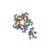
|
|---|---|
| 1 | x 60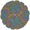
|
| 2 |
|
| 3 | x 5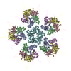
|
| 4 | x 6
|
| 5 | 
|
| Symmetry | Point symmetry: (Schoenflies symbol: I (icosahedral)) |
-Components
| #1: Protein | Mass: 55619.023 Da / Num. of mol.: 6 / Source method: isolated from a natural source Details: BPV was purified from cow warts on a CsCl gradient (Li et al (1998) J Virol 72: 2160-2167) Source: (natural) Bovine papillomavirus type 1 / Strain: BPV type 1 / References: UniProt: P03103 |
|---|
-Experimental details
-Experiment
| Experiment | Method: ELECTRON MICROSCOPY |
|---|---|
| EM experiment | Aggregation state: PARTICLE / 3D reconstruction method: single particle reconstruction |
- Sample preparation
| Component | Name: BOVINE PAPILLOMAVIRUS TYPE 1 (BPV1), FULL VIRION / Type: VIRUS |
|---|---|
| Details of virus | Host category: VERTEBRATES / Isolate: SEROTYPE / Type: VIRION |
| Natural host | Organism: Bos taurus |
| Buffer solution | Name: 20MM TRIS PH 6.2, 100MM NACL, 0.5MM CACL2 / pH: 6.2 / Details: 20MM TRIS PH 6.2, 100MM NACL, 0.5MM CACL2 |
| Specimen | Conc.: 1.5 mg/ml / Embedding applied: NO / Shadowing applied: NO / Staining applied: NO / Vitrification applied: YES |
| Specimen support | Details: C-FLAT CF-1/2-4C |
| Vitrification | Instrument: HOMEMADE PLUNGER / Cryogen name: ETHANE / Details: VITRIFICATION CARRIED OUT IN COLD ROOM. |
- Electron microscopy imaging
| Experimental equipment |  Model: Tecnai F30 / Image courtesy: FEI Company |
|---|---|
| Microscopy | Model: FEI TECNAI F30 / Date: Dec 7, 2008 / Details: OBJ APERTURE CUTOFF AT 2.4A |
| Electron gun | Electron source: FIELD EMISSION GUN / Accelerating voltage: 300 kV / Illumination mode: FLOOD BEAM |
| Electron lens | Mode: BRIGHT FIELDBright-field microscopy / Nominal magnification: 59000 X / Calibrated magnification: 56588 X / Nominal defocus max: 2900 nm / Nominal defocus min: 1800 nm / Cs: 2 mm |
| Specimen holder | Temperature: 77 K / Tilt angle max: 0 ° / Tilt angle min: 0 ° |
| Image recording | Electron dose: 25 e/Å2 / Film or detector model: KODAK SO-163 FILM |
| Image scans | Num. digital images: 49 |
- Processing
| EM software |
| ||||||||||||
|---|---|---|---|---|---|---|---|---|---|---|---|---|---|
| CTF correction | Details: CTFILT3 WITH INDIVIDUAL PARTICLE ADJUSTMENT | ||||||||||||
| Symmetry | Point symmetry: I (icosahedral) | ||||||||||||
| 3D reconstruction | Method: CENTRAL SECTIONS / Resolution: 4.2 Å / Num. of particles: 3997 / Nominal pixel size: 1.186 Å / Actual pixel size: 1.237 Å Magnification calibration: ATOMIC PENTAMER MODEL, MAXIMIZED REAL-SPACE CORRELATION USING SITUS Details: EWALD SPHERE CORRECTION / Symmetry type: POINT | ||||||||||||
| Atomic model building | B value: 131 / Protocol: RIGID BODY FIT / Space: RECIPROCAL Target criteria: MLHL TARGET WITH AMPL. AND PHASE PROBABILITY DISTRIBUTION Details: METHOD--RIGID BODY DOCKING, THEN COORDINATE REFINEMENT IN CNS REFINEMENT PROTOCOL--SEE 3D-FITTING DETAILS | ||||||||||||
| Atomic model building | PDB-ID: 1DZL Details: HOMOLOGY MODEL 1DZL | ||||||||||||
| Refinement | Highest resolution: 4.2 Å | ||||||||||||
| Displacement parameters | Baniso 11: 131 Å2 | ||||||||||||
| Refinement step | Cycle: LAST / Highest resolution: 4.2 Å
|

