 Movie
Movie Controller

[English] 日本語
 Yorodumi
- PDB-4c4q: Cryo-EM map of the CSFV IRES in complex with the small ribosomal ... -
+ Open data

- Basic information
| Entry | Database: PDB / ID: 4c4q | |||||||||
|---|---|---|---|---|---|---|---|---|---|---|
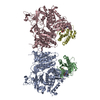
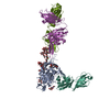
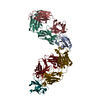
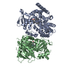
| Title | Cryo-EM map of the CSFV IRES in complex with the small ribosomal 40S subunit and DHX29 | |||||||||
 Components Components | INTERNAL RIBOSOMAL ENTRY SITE | |||||||||
 Keywords Keywords | RNA / INTERNAL RIBOSOMAL ENTRY SITE / 5'-END INDEPENDENT INITIATION / HCV-LIKE IRES | |||||||||
| Function / homology | : / RNA / RNA (> 10) / RNA (> 100) Function and homology information Function and homology information | |||||||||
| Biological species |  CLASSICAL SWINE FEVER VIRUS CLASSICAL SWINE FEVER VIRUS | |||||||||
| Method | ELECTRON MICROSCOPY / single particle reconstruction / cryo EM / Resolution: 8.5 Å | |||||||||
 Authors Authors | Hashem, Y. / desGeorges, A. / Dhote, V. / Langlois, R. / Liao, H.Y. / Grassucci, R.A. / Pestova, T.V. / Hellen, C.U.T. / Frank, J. | |||||||||
 Citation Citation | Journal: Nature / Year: 2013 Title: Hepatitis-C-virus-like internal ribosome entry sites displace eIF3 to gain access to the 40S subunit. Authors: Yaser Hashem / Amedee des Georges / Vidya Dhote / Robert Langlois / Hstau Y Liao / Robert A Grassucci / Tatyana V Pestova / Christopher U T Hellen / Joachim Frank /  Abstract: Hepatitis C virus (HCV) and classical swine fever virus (CSFV) messenger RNAs contain related (HCV-like) internal ribosome entry sites (IRESs) that promote 5'-end independent initiation of ...Hepatitis C virus (HCV) and classical swine fever virus (CSFV) messenger RNAs contain related (HCV-like) internal ribosome entry sites (IRESs) that promote 5'-end independent initiation of translation, requiring only a subset of the eukaryotic initiation factors (eIFs) needed for canonical initiation on cellular mRNAs. Initiation on HCV-like IRESs relies on their specific interaction with the 40S subunit, which places the initiation codon into the P site, where it directly base-pairs with eIF2-bound initiator methionyl transfer RNA to form a 48S initiation complex. However, all HCV-like IRESs also specifically interact with eIF3 (refs 2, 5-7, 9-12), but the role of this interaction in IRES-mediated initiation has remained unknown. During canonical initiation, eIF3 binds to the 40S subunit as a component of the 43S pre-initiation complex, and comparison of the ribosomal positions of eIF3 and the HCV IRES revealed that they overlap, so that their rearrangement would be required for formation of ribosomal complexes containing both components. Here we present a cryo-electron microscopy reconstruction of a 40S ribosomal complex containing eIF3 and the CSFV IRES. Remarkably, although the position and interactions of the CSFV IRES with the 40S subunit in this complex are similar to those of the HCV IRES in the 40S-IRES binary complex, eIF3 is completely displaced from its ribosomal position in the 43S complex, and instead interacts through its ribosome-binding surface exclusively with the apical region of domain III of the IRES. Our results suggest a role for the specific interaction of HCV-like IRESs with eIF3 in preventing ribosomal association of eIF3, which could serve two purposes: relieving the competition between the IRES and eIF3 for a common binding site on the 40S subunit, and reducing formation of 43S complexes, thereby favouring translation of viral mRNAs. | |||||||||
| History |
|
- Structure visualization
| Movie |
Movie viewer |
|---|---|
| Structure viewer | Molecule: MolmilJmol/JSmol |
- Downloads & links
-Download
| PDBx/mmCIF format | 4c4q.cif.gz | 140.7 KB | Display | PDBx/mmCIF format |
|---|---|---|---|---|
| PDB format | pdb4c4q.ent.gz | 86.2 KB | Display | PDB format |
| PDBx/mmJSON format | 4c4q.json.gz | Tree view | PDBx/mmJSON format | |
| Others |  Other downloads Other downloads |
-Validation report
| Summary document | 4c4q_validation.pdf.gz | 827.6 KB | Display | wwPDB validaton report |
|---|---|---|---|---|
| Full document | 4c4q_full_validation.pdf.gz | 1021.4 KB | Display | |
| Data in XML | 4c4q_validation.xml.gz | 33.9 KB | Display | |
| Data in CIF | 4c4q_validation.cif.gz | 45.2 KB | Display | |
| Arichive directory | https://data.pdbj.org/pub/pdb/validation_reports/c4/4c4qftp://data.pdbj.org/pub/pdb/validation_reports/c4/4c4q | HTTPS FTP |
-Related structure data
| Related structure data | 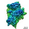 2450MC 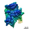 2451C M: map data used to model this data C: citing same article ( |
|---|---|
| Similar structure data |

-Links
 PDBj
PDBj


- Assembly
| Deposited unit | 
|
|---|---|
| 1 |
|
-Components
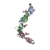
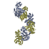
| #1: RNA chain | Mass: 75351.680 Da / Num. of mol.: 1 / Fragment: DOMAIN III OF CSFV IRES, RESIDUES 128-360 / Source method: obtained synthetically Details: AS DOMAIN II IS TRUNCATED, THE IRES STRUCTURED BODY CONSISTS MAINLY ON DOMAINS III (RESIDUES 128-360) Source: (synth.) CLASSICAL SWINE FEVER VIRUS / References: GenBank: J04358 |
|---|
-Experimental details
-Experiment
| Experiment | Method: ELECTRON MICROSCOPY |
|---|---|
| EM experiment | Aggregation state: PARTICLE / 3D reconstruction method: single particle reconstruction |
- Sample preparation
| Component | Name: CSFV IRES TRUNCATED FROM DOMAIN II, IN COMPLEX WITH THE RABBIT SMALL RIBOSOMAL 40S SUBUNIT AND TO DHX29 Type: RIBOSOME |
|---|---|
| Buffer solution | Name: 20 MM TRIS, 75 MM KCL, 5MM MG, 2 MM DTT AND 0.25 MM SPERMIDINE pH: 7.5 Details: 20 MM TRIS, 75 MM KCL, 5MM MG, 2 MM DTT AND 0.25 MM SPERMIDINE |
| Specimen | Conc.: 0.1 mg/ml / Embedding applied: NO / Shadowing applied: NO / Staining applied: NO / Vitrification applied: YES |
| Specimen support | Details: CARBON |
| Vitrification | Instrument: FEI VITROBOT MARK II / Cryogen name: ETHANE Details: VITRIFICATION 1 -- CRYOGEN- ETHANE, HUMIDITY- 100, TEMPERATURE- 120, INSTRUMENT- FEI VITROBOT MARK II, METHOD- 30 SECONDS WAITING AFTER SAMPLE DEPOSITION ON THE GRID, BLOTTING FOR SECONDS BEFORE PLUNGING, |
- Electron microscopy imaging
| Experimental equipment |  Model: Tecnai F20 / Image courtesy: FEI Company |
|---|---|
| Microscopy | Model: FEI TECNAI F20 / Date: Feb 1, 2013 |
| Electron gun | Electron source:  FIELD EMISSION GUN / Accelerating voltage: 120 kV / Illumination mode: FLOOD BEAM FIELD EMISSION GUN / Accelerating voltage: 120 kV / Illumination mode: FLOOD BEAM |
| Electron lens | Mode: BRIGHT FIELD / Calibrated magnification: 51570 X / Nominal defocus max: 4000 nm / Nominal defocus min: 1000 nm / Cs: 2.26 mm |
| Specimen holder | Temperature: 110 K |
| Image recording | Electron dose: 12 e/Å2 / Film or detector model: GATAN ULTRASCAN 4000 (4k x 4k) |
| Image scans | Num. digital images: 2000 |
| Radiation wavelength | Relative weight: 1 |
- Processing
| EM software |
| ||||||||||||
|---|---|---|---|---|---|---|---|---|---|---|---|---|---|
| CTF correction | Details: EACH PARTICLE | ||||||||||||
| Symmetry | Point symmetry: C1 (asymmetric) | ||||||||||||
| 3D reconstruction | Method: TEMPLATE MATCHING / Resolution: 8.5 Å / Num. of particles: 72900 / Actual pixel size: 2.245 Å Details: SUBMISSION BASED ON EXPERIMENTAL DATA FROM EMDB EMD-2450. (DEPOSITION ID: 11933) Symmetry type: POINT | ||||||||||||
| Atomic model building | Protocol: OTHER / Space: RECIPROCAL / Details: REFINEMENT PROTOCOL--X-RAY | ||||||||||||
| Atomic model building | PDB-ID: 2XZM 2xzm Accession code: 2XZM / Source name: PDB / Type: experimental model | ||||||||||||
| Refinement | Highest resolution: 8.5 Å | ||||||||||||
| Refinement step | Cycle: LAST / Highest resolution: 8.5 Å
|
