 Movie
Movie Controller


+ Open data

- Basic information
| Entry | Database: PDB / ID: 1m2o | ||||||
|---|---|---|---|---|---|---|---|
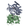
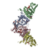
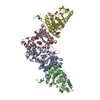
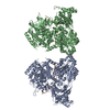
| Title | Crystal Structure of the Sec23-Sar1 complex | ||||||
 Components Components |
| ||||||
 Keywords Keywords | PROTEIN TRANSPORT/SIGNALING PROTEIN /  zinc-finger / beta barrel / vWA domain / gelsolin domain / PROTEIN TRANSPORT-SIGNALING PROTEIN COMPLEX zinc-finger / beta barrel / vWA domain / gelsolin domain / PROTEIN TRANSPORT-SIGNALING PROTEIN COMPLEX | ||||||
| Function / homology |  Function and homology information Function and homology informationAntigen Presentation: Folding, assembly and peptide loading of class I MHC / Cargo concentration in the ER / regulation of COPII vesicle coating / positive regulation of ER to Golgi vesicle-mediated transport / mitochondria-associated endoplasmic reticulum membrane contact site / COPII-mediated vesicle transport / nuclear envelope organization / COPII-coated vesicle cargo loading / vesicle organization / COPII vesicle coat ...Antigen Presentation: Folding, assembly and peptide loading of class I MHC / Cargo concentration in the ER / regulation of COPII vesicle coating / positive regulation of ER to Golgi vesicle-mediated transport / mitochondria-associated endoplasmic reticulum membrane contact site / COPII-mediated vesicle transport / nuclear envelope organization / COPII-coated vesicle cargo loading / vesicle organization / COPII vesicle coat / positive regulation of protein exit from endoplasmic reticulum / membrane organization / mitochondrial fission / mitochondrial membrane organization / reticulophagy / endoplasmic reticulum to Golgi vesicle-mediated transport / endoplasmic reticulum exit site / GTPase activator activity / macroautophagy / Hydrolases; Acting on acid anhydrides; Acting on GTP to facilitate cellular and subcellular movement / intracellular protein transport / Golgi membrane / GTPase activity / GTP binding / endoplasmic reticulum membrane / endoplasmic reticulum / mitochondrion / zinc ion bindingSimilarity search - Function | ||||||
| Biological species |  Saccharomyces cerevisiae (brewer's yeast) Saccharomyces cerevisiae (brewer's yeast) | ||||||
| Method | X-RAY DIFFRACTION / SYNCHROTRON / MAD / Resolution: 2.5 Å | ||||||
 Authors Authors | Bi, X. / Corpina, R.A. / Goldberg, J. | ||||||
 Citation Citation | Journal: Nature / Year: 2002 Title: Structure of the Sec23/24-Sar1 pre-budding complex of the COPII vesicle coat Authors: Bi, X. / Corpina, R.A. / Goldberg, J. | ||||||
| History |
|
- Structure visualization
| Structure viewer | Molecule: MolmilJmol/JSmol |
|---|
- Downloads & links
-Download
| PDBx/mmCIF format | 1m2o.cif.gz | 360.2 KB | Display | PDBx/mmCIF format |
|---|---|---|---|---|
| PDB format | pdb1m2o.ent.gz | 287.2 KB | Display | PDB format |
| PDBx/mmJSON format | 1m2o.json.gz | Tree view | PDBx/mmJSON format | |
| Others |  Other downloads Other downloads |
-Validation report
| Arichive directory | https://data.pdbj.org/pub/pdb/validation_reports/m2/1m2oftp://data.pdbj.org/pub/pdb/validation_reports/m2/1m2o | HTTPS FTP |
|---|
-Related structure data


-Links
 PDBj
PDBj


- Assembly
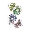
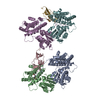
| Deposited unit | 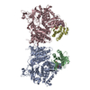
| ||||||||||
|---|---|---|---|---|---|---|---|---|---|---|---|
| 1 |
| ||||||||||
| 2 | 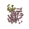
| ||||||||||
| 3 | 
| ||||||||||
| Unit cell |
|
-Components
-Protein , 2 types, 4 molecules ACBD
| #1: Protein | Protein targeting / Sec23 / Sec23p / cytoplasmic GTPase-activating protein / Sec23 COPII-coat protein Mass: 85463.242 Da / Num. of mol.: 2 Source method: isolated from a genetically manipulated source Source: (gene. exp.) Saccharomyces cerevisiae (brewer's yeast)Gene: Sec23 / Production host:  Escherichia coli (E. coli) / References: UniProt: P15303 Escherichia coli (E. coli) / References: UniProt: P15303#2: Protein | Mass: 21472.564 Da / Num. of mol.: 2 Source method: isolated from a genetically manipulated source Source: (gene. exp.) Saccharomyces cerevisiae (brewer's yeast)Gene: Sar1 / Production host: Escherichia coli (E. coli) / References: UniProt: P20606 |
|---|
-Non-polymers , 4 types, 177 molecules 


| #3: Chemical |  Mass: 65.409 Da / Num. of mol.: 2 / Source method: obtained synthetically / Formula: Zn Mass: 65.409 Da / Num. of mol.: 2 / Source method: obtained synthetically / Formula: Zn#4: Chemical |  Mass: 24.305 Da / Num. of mol.: 2 / Source method: obtained synthetically / Formula: Mg Mass: 24.305 Da / Num. of mol.: 2 / Source method: obtained synthetically / Formula: Mg#5: Chemical | 5'-Guanylyl imidodiphosphate Mass: 522.196 Da / Num. of mol.: 2 / Source method: obtained synthetically / Formula: C10H17N6O13P3 Mass: 522.196 Da / Num. of mol.: 2 / Source method: obtained synthetically / Formula: C10H17N6O13P3Comment: GppNHp, GMPPNP, energy-carrying molecule analogue*YM #6: Water | ChemComp-HOH / | WaterMass: 18.015 Da / Num. of mol.: 171 / Source method: isolated from a natural source / Formula: H2O |
|---|
-Experimental details
-Experiment
| Experiment | Method: X-RAY DIFFRACTION / Number of used crystals: 1 |
|---|
- Sample preparation
| Crystal | Density Matthews: 2.26 Å3/Da / Density % sol: 45.67 % | ||||||||||||||||||||||||||||||||||||
|---|---|---|---|---|---|---|---|---|---|---|---|---|---|---|---|---|---|---|---|---|---|---|---|---|---|---|---|---|---|---|---|---|---|---|---|---|---|
| Crystal grow | Temperature: 270 K / Method: vapor diffusion, hanging drop / pH: 6.5 Details: 12% (w/v) PEG 1500, 10% (w/v) isopropanol, 0.2 M ammonium acetate, 50 mM MES, pH 6.5, VAPOR DIFFUSION, HANGING DROP at 270K | ||||||||||||||||||||||||||||||||||||
| Crystal grow | *PLUS Temperature: 22 ℃ | ||||||||||||||||||||||||||||||||||||
| Components of the solutions | *PLUS
|
-Data collection
| Diffraction | Mean temperature: 100 K |
|---|---|
| Diffraction source | Source: SYNCHROTRON / Site: CHESS  / Beamline: F2 / Wavelength: 1 Å / Beamline: F2 / Wavelength: 1 Å |
| Detector | Type: KODAK / Detector: IMAGE PLATE / Date: Nov 5, 2001 |
| Radiation | Monochromator: CHESS F-2 / Protocol: SINGLE WAVELENGTH / Monochromatic (M) / Laue (L): M / Scattering type: x-ray |
| Radiation wavelength | Wavelength: 1 Å / Relative weight: 1 |
| Reflection | Resolution: 2.5→25 Å / Num. all: 68753 / Num. obs: 63872 / % possible obs: 92.9 % / Observed criterion σ(I): -3 / Biso Wilson estimate: 21.4 Å2 / Rmerge(I) obs: 0.046 |
| Reflection shell | Resolution: 2.5→2.66 Å |
| Reflection | *PLUS Lowest resolution: 25 Å / Num. measured all: 289977 / Rmerge(I) obs: 0.046 |
| Reflection shell | *PLUS % possible obs: 79.9 % / Rmerge(I) obs: 0.237 |
- Processing
| Software |
| ||||||||||||||||||||||||||||||||||||
|---|---|---|---|---|---|---|---|---|---|---|---|---|---|---|---|---|---|---|---|---|---|---|---|---|---|---|---|---|---|---|---|---|---|---|---|---|---|
| Refinement | Method to determine structure: MAD / Resolution: 2.5→23.85 Å / Rfactor Rfree error: 0.005 / Isotropic thermal model: RESTRAINED / Cross valid method: THROUGHOUT / σ(F): 0 / σ(I): 0 / Stereochemistry target values: Engh & Huber
| ||||||||||||||||||||||||||||||||||||
| Solvent computation | Solvent model: FLAT MODEL / Bsol: 23.6345 Å2 / ksol: 0.312224 e/Å3 | ||||||||||||||||||||||||||||||||||||
| Displacement parameters | Biso mean: 43.3 Å2
| ||||||||||||||||||||||||||||||||||||
| Refine analyze | Luzzati coordinate error free: 0.45 Å / Luzzati sigma a free: 0.42 Å | ||||||||||||||||||||||||||||||||||||
| Refinement step | Cycle: LAST / Resolution: 2.5→23.85 Å
| ||||||||||||||||||||||||||||||||||||
| Refine LS restraints |
| ||||||||||||||||||||||||||||||||||||
| LS refinement shell | Resolution: 2.5→2.66 Å / Rfactor Rfree error: 0.018 / Total num. of bins used: 6
| ||||||||||||||||||||||||||||||||||||
| Xplor file |
| ||||||||||||||||||||||||||||||||||||
| Refinement | *PLUS % reflection Rfree: 5 % / Rfactor all: 0.238 / Rfactor obs: 0.238 / Rfactor Rfree: 0.295 / Rfactor Rwork: 0.238 | ||||||||||||||||||||||||||||||||||||
| Solvent computation | *PLUS | ||||||||||||||||||||||||||||||||||||
| Displacement parameters | *PLUS | ||||||||||||||||||||||||||||||||||||
| Refine LS restraints | *PLUS
| ||||||||||||||||||||||||||||||||||||
| LS refinement shell | *PLUS Rfactor Rfree: 0.368 / Rfactor Rwork: 0.282 |
