 Movie
Movie Controller


+ Open data

- Basic information
| Entry | Database: PDB / ID: 3j5v | ||||||
|---|---|---|---|---|---|---|---|
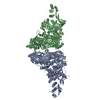
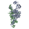
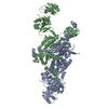
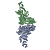
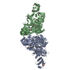
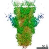
| Title | PhuZ201 filament | ||||||
 Components Components | PhuZ201 subunit | ||||||
 Keywords Keywords | VIRAL PROTEIN / PhuZ / tubulin / FtsZ / filament / bacteriophage / cytoskeleton / bacteriophage centering function | ||||||
| Function / homology | establishment of localization / Tubulin/FtsZ, GTPase domain superfamily / Hydrolases; Acting on acid anhydrides; Acting on GTP to facilitate cellular and subcellular movement / host cell cytoplasm / GTPase activity / GTP binding / identical protein binding / GUANOSINE-5'-DIPHOSPHATE / Phage tubulin-like protein Function and homology information Function and homology information | ||||||
| Biological species |  Pseudomonas phage 201phi2-1 (virus) Pseudomonas phage 201phi2-1 (virus) | ||||||
| Method | ELECTRON MICROSCOPY / helical reconstruction / cryo EM / Resolution: 7.1 Å | ||||||
 Authors Authors | Zehr, E.A. | ||||||
 Citation Citation | Journal: Structure / Year: 2014 Title: The structure and assembly mechanism of a novel three-stranded tubulin filament that centers phage DNA. Authors: Elena A Zehr / James A Kraemer / Marcella L Erb / Joanna K C Coker / Elizabeth A Montabana / Joe Pogliano / David A Agard /  Abstract: Tubulins are a universally conserved protein superfamily that carry out diverse biological roles by assembling filaments with very different architectures. The underlying basis of this structural ...Tubulins are a universally conserved protein superfamily that carry out diverse biological roles by assembling filaments with very different architectures. The underlying basis of this structural diversity is poorly understood. Here, we determine a 7.1 Å cryo-electron microscopy reconstruction of the bacteriophage-encoded PhuZ filament and provide molecular-level insight into its cooperative assembly mechanism. The PhuZ family of tubulins is required to actively center the phage within infected host cells, facilitating efficient phage replication. Our reconstruction and derived model reveal the first example of a three-stranded tubulin filament. We show that the elongated C-terminal tail simultaneously stabilizes both longitudinal and lateral interactions, which in turn define filament architecture. Identified interaction surfaces are conserved within the PhuZ family, and their mutagenesis compromises polymerization in vitro and in vivo. Combining kinetic modeling of PhuZ filament assembly and structural data, we suggest a common filament structure and assembly mechanism for the PhuZ family of tubulins. | ||||||
| History |
|
- Structure visualization
| Movie |
Movie viewer |
|---|---|
| Structure viewer | Molecule: MolmilJmol/JSmol |
- Downloads & links
-Download
| PDBx/mmCIF format | 3j5v.cif.gz | 77 KB | Display | PDBx/mmCIF format |
|---|---|---|---|---|
| PDB format | pdb3j5v.ent.gz | 56.1 KB | Display | PDB format |
| PDBx/mmJSON format | 3j5v.json.gz | Tree view | PDBx/mmJSON format | |
| Others |  Other downloads Other downloads |
-Validation report
| Summary document | 3j5v_validation.pdf.gz | 874.9 KB | Display | wwPDB validaton report |
|---|---|---|---|---|
| Full document | 3j5v_full_validation.pdf.gz | 873.4 KB | Display | |
| Data in XML | 3j5v_validation.xml.gz | 15.9 KB | Display | |
| Data in CIF | 3j5v_validation.cif.gz | 22 KB | Display | |
| Arichive directory | https://data.pdbj.org/pub/pdb/validation_reports/j5/3j5vftp://data.pdbj.org/pub/pdb/validation_reports/j5/3j5v | HTTPS FTP |
-Related structure data
| Related structure data | 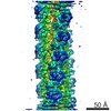 5783MC M: map data used to model this data C: citing same article ( |
|---|---|
| Similar structure data |

-Links
 PDBj
PDBj


- Assembly
| Deposited unit | 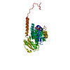
|
|---|---|
| 1 | x 9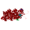
|
| 2 |
|
| 3 | 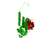
|
| Symmetry | Helical symmetry: (Circular symmetry: 1 / Dyad axis: no / N subunits divisor: 1 / Num. of operations: 9 / Rise per n subunits: 14.42 Å / Rotation per n subunits: -116.44 °) |
-Components
| #1: Protein | Mass: 35100.004 Da / Num. of mol.: 1 Source method: isolated from a genetically manipulated source Source: (gene. exp.) Pseudomonas phage 201phi2-1 (virus) / Gene: 201phi2-1p059 / Plasmid: pET28a(+) / Production host:  |
|---|---|
| #2: Chemical | ChemComp-GDP /   Type: RNA linking / Mass: 443.201 Da / Num. of mol.: 1 / Source method: obtained synthetically / Formula: C10H15N5O11P2 / Comment: GDP, energy-carrying molecule*YM Type: RNA linking / Mass: 443.201 Da / Num. of mol.: 1 / Source method: obtained synthetically / Formula: C10H15N5O11P2 / Comment: GDP, energy-carrying molecule*YM |
| #3: Chemical | ChemComp-MG /   Mass: 24.305 Da / Num. of mol.: 1 / Source method: obtained synthetically / Formula: Mg Mass: 24.305 Da / Num. of mol.: 1 / Source method: obtained synthetically / Formula: Mg |
-Experimental details
-Experiment
| Experiment | Method: ELECTRON MICROSCOPY |
|---|---|
| EM experiment | Aggregation state: FILAMENT / 3D reconstruction method: helical reconstruction |
- Sample preparation
| Component | Name: PhuZ filament / Type: COMPLEX |
|---|---|
| Molecular weight | Value: 0.034621 MDa / Experimental value: NO |
| Buffer solution | Name: 250 mM KCl, 50 mM HEPES pH 7.4, 1 mM MgCl2, 10% glycerol, 1mM DTT pH: 8 Details: 250 mM KCl, 50 mM HEPES pH 7.4, 1 mM MgCl2, 10% glycerol, 1mM DTT |
| Specimen | Conc.: 0.7 mg/ml / Embedding applied: NO / Shadowing applied: NO / Staining applied: NO / Vitrification applied: YES |
| Specimen support | Details: C-FLAT holey carbon grid, 200 mesh copper grid |
| Vitrification | Instrument: FEI VITROBOT MARK III / Cryogen name: ETHANE / Humidity: 90 % Details: Blotted 2 seconds before plunging in liquid ethane (FEI VITROBOT MARK III) |
- Electron microscopy imaging
| Experimental equipment |  Model: Tecnai F20 / Image courtesy: FEI Company |
|---|---|
| Microscopy | Model: FEI TECNAI F20 / Date: Nov 21, 2011 |
| Electron gun | Electron source:  FIELD EMISSION GUN / Accelerating voltage: 200 kV / Illumination mode: FLOOD BEAM FIELD EMISSION GUN / Accelerating voltage: 200 kV / Illumination mode: FLOOD BEAM |
| Electron lens | Mode: BRIGHT FIELD / Nominal magnification: 62000 X / Nominal defocus max: 2500 nm / Nominal defocus min: 700 nm / Cs: 2.2 mm |
| Image recording | Electron dose: 25 e/Å2 / Film or detector model: TVIPS TEMCAM-F816 (8k x 8k) |
| Image scans | Num. digital images: 461 |
- Processing
| EM software |
| ||||||||||||
|---|---|---|---|---|---|---|---|---|---|---|---|---|---|
| CTF correction | Details: Whole micrograph | ||||||||||||
| Helical symmerty | Angular rotation/subunit: -116.44 ° / Axial rise/subunit: 14.42 Å / Axial symmetry: C1 | ||||||||||||
| 3D reconstruction | Method: Helical reconstruction / Resolution: 7.1 Å / Num. of particles: 23243 / Nominal pixel size: 1.20398 Å Details: A modified version of SPIDER program was used for the reconstruction. Helical symmetry search was performed with hsearch_lorentz. Symmetry type: HELICAL | ||||||||||||
| Atomic model building | Protocol: FLEXIBLE FIT / Space: REAL / Target criteria: cross-correlation Details: METHOD--Flexible fitting DETAILS--Flexible fitting applied to C-terminus (residues 273-315) | ||||||||||||
| Atomic model building | PDB-ID: 3R4V Accession code: 3R4V / Source name: PDB / Type: experimental model | ||||||||||||
| Refinement | Highest resolution: 7.1 Å | ||||||||||||
| Refinement step | Cycle: LAST / Highest resolution: 7.1 Å
|
