 Movie
Movie Controller

[English] 日本語
 Yorodumi
- PDB-6acx: Crystal structure of Mycobacterium smegmatis Mfd in complex with ... -
+ Open data

- Basic information
| Entry | Database: PDB / ID: 6acx | ||||||
|---|---|---|---|---|---|---|---|
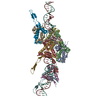
| Title | Crystal structure of Mycobacterium smegmatis Mfd in complex with ADP + Pi at 3.5 A resolution. | ||||||
 Components Components | Mycobacterium smegmatis Mfd | ||||||
 Keywords Keywords |  HYDROLASE / Transcription repair coupling factor / Mfd / Transcription regulation / Transcription Coupled Nucleotide Excision Repair. HYDROLASE / Transcription repair coupling factor / Mfd / Transcription regulation / Transcription Coupled Nucleotide Excision Repair. | ||||||
| Function / homology |  Function and homology information Function and homology informationtranscription-coupled nucleotide-excision repair, DNA damage recognition / helicase activity / Hydrolases; Acting on acid anhydrides; Acting on acid anhydrides to facilitate cellular and subcellular movement / damaged DNA binding / hydrolase activity / regulation of DNA-templated transcription / ATP binding / cytoplasmSimilarity search - Function | ||||||
| Biological species |  Mycolicibacterium smegmatis MC2 155 (bacteria) Mycolicibacterium smegmatis MC2 155 (bacteria) | ||||||
| Method | X-RAY DIFFRACTION / MOLECULAR REPLACEMENT / Resolution: 3.5 Å | ||||||
 Authors Authors | Putta, S. / Fox, G.C. / Walsh, M.A. / Rao, D.N. / Nagaraja, V. / Natesh, R. | ||||||
| Funding support |  India, 1items India, 1items
| ||||||
 Citation Citation | Journal: To Be Published Title: Structural basis for nucleotide-mediated remodelling mechanism of Mycobacterium Mfd Authors: Putta, S. / Prabha, S. / Bhat, V. / Fox, G.C. / Walsh, M.A. / Rao, D.N. / Nagaraja, V. / Natesh, R. | ||||||
| History |
|
- Structure visualization
| Structure viewer | Molecule: MolmilJmol/JSmol |
|---|
- Downloads & links
-Download
| PDBx/mmCIF format | 6acx.cif.gz | 453.4 KB | Display | PDBx/mmCIF format |
|---|---|---|---|---|
| PDB format | pdb6acx.ent.gz | 354.4 KB | Display | PDB format |
| PDBx/mmJSON format | 6acx.json.gz | Tree view | PDBx/mmJSON format | |
| Others |  Other downloads Other downloads |
-Validation report
| Arichive directory | https://data.pdbj.org/pub/pdb/validation_reports/ac/6acxftp://data.pdbj.org/pub/pdb/validation_reports/ac/6acx | HTTPS FTP |
|---|
-Related structure data
| Related structure data |  9614C  6ac6C  6ac8SC  6acaC C: citing same article ( S: Starting model for refinement |
|---|---|
| Similar structure data |


-Links
 PDBj
PDBj- Assembly
| Deposited unit | 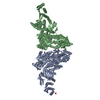
| ||||||||
|---|---|---|---|---|---|---|---|---|---|
| 1 |
| ||||||||
| 2 | 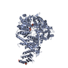
| ||||||||
| 3 | 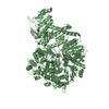
| ||||||||
| Unit cell |
|
-Components
| #1: Protein | Mass: 133438.484 Da / Num. of mol.: 2 Source method: isolated from a genetically manipulated source Details: Cloned from Genomic DNA of Mycobacterium smegmatis Source: (gene. exp.) Mycolicibacterium smegmatis MC2 155 (bacteria)Strain: MC2 155 / Gene: mfd / Plasmid: pETMsMfd Details (production host): Amplified from Genomic DNA, Cloned into pET28a Production host: Escherichia coli (E. coli) / Strain (production host): Rosetta (DE3)References: UniProt: I7G7M2, UniProt: A0R3C5*PLUS, Hydrolases; Acting on acid anhydrides; Acting on acid anhydrides to facilitate cellular and subcellular movement#2: Chemical | ChemComp-SO4 / Sulfate  Mass: 96.063 Da / Num. of mol.: 4 / Source method: obtained synthetically / Formula: SO4 Mass: 96.063 Da / Num. of mol.: 4 / Source method: obtained synthetically / Formula: SO4#3: Chemical | ChemComp-PO4 / | Phosphate  Mass: 94.971 Da / Num. of mol.: 1 / Source method: obtained synthetically / Formula: PO4 Mass: 94.971 Da / Num. of mol.: 1 / Source method: obtained synthetically / Formula: PO4#4: Chemical | Adenosine diphosphate  Mass: 427.201 Da / Num. of mol.: 2 / Source method: obtained synthetically / Formula: C10H15N5O10P2 / Comment: ADP, energy-carrying molecule*YM Mass: 427.201 Da / Num. of mol.: 2 / Source method: obtained synthetically / Formula: C10H15N5O10P2 / Comment: ADP, energy-carrying molecule*YM#5: Water | ChemComp-HOH / | Water Mass: 18.015 Da / Num. of mol.: 105 / Source method: isolated from a natural source / Formula: H2O Mass: 18.015 Da / Num. of mol.: 105 / Source method: isolated from a natural source / Formula: H2O |
|---|
-Experimental details
-Experiment
| Experiment | Method: X-RAY DIFFRACTION / Number of used crystals: 1 |
|---|
- Sample preparation
| Crystal | Density Matthews: 2.7 Å3/Da / Density % sol: 54.4 % Description: THE ENTRY CONTAINS FRIEDEL PAIRS IN I/F_PLUS/MINUS COLUMNS. |
|---|---|
| Crystal grow | Temperature: 295 K / Method: vapor diffusion, hanging drop / pH: 7.2 Details: 100mM HEPES sodium pH 7.2, 0.2M Na2So4, 20% PEG3350 PH range: 7.2 - 7.5 / Temp details: Room temperatur |
-Data collection
| Diffraction | Mean temperature: 100 K / Ambient temp details: Oxford Cryo Strem |
|---|---|
| Diffraction source | Source: ROTATING ANODE / Type: BRUKER AXS MICROSTAR-H / Wavelength: 1.5418 Å |
| Detector | Type: MAR scanner 345 mm plate / Detector: IMAGE PLATE / Date: Mar 1, 2017 / Details: HELIOS MX |
| Radiation | Monochromator: HELIOS MX mirrors / Protocol: SINGLE WAVELENGTH / Monochromatic (M) / Laue (L): M / Scattering type: x-ray |
| Radiation wavelength | Wavelength: 1.5418 Å / Relative weight: 1 |
| Reflection | Resolution: 3.5→31.73 Å / Num. obs: 35989 / % possible obs: 96.8 % / Redundancy: 5.1 % / Biso Wilson estimate: 43.755 Å2 / CC1/2: 0.982 / Rmerge(I) obs: 0.2 / Rpim(I) all: 0.104 / Rrim(I) all: 0.245 / Rsym value: 0.22 / Net I/σ(I): 6.3 |
| Reflection shell | Resolution: 3.5→3.66 Å / Redundancy: 4.9 % / Rmerge(I) obs: 0.594 / Mean I/σ(I) obs: 2.4 / Num. unique obs: 4329 / CC1/2: 0.353 / Rpim(I) all: 0.305 / Rrim(I) all: 0.711 / Rsym value: 0.637 / % possible all: 97.1 |
- Processing
| Software |
| ||||||||||||||||||||||||||||||||||||||||||||||||||||||||||||||||||||||||||||||||||||||||||||||||||
|---|---|---|---|---|---|---|---|---|---|---|---|---|---|---|---|---|---|---|---|---|---|---|---|---|---|---|---|---|---|---|---|---|---|---|---|---|---|---|---|---|---|---|---|---|---|---|---|---|---|---|---|---|---|---|---|---|---|---|---|---|---|---|---|---|---|---|---|---|---|---|---|---|---|---|---|---|---|---|---|---|---|---|---|---|---|---|---|---|---|---|---|---|---|---|---|---|---|---|---|
| Refinement | Method to determine structure: MOLECULAR REPLACEMENT Starting model: 6AC8 Resolution: 3.5→30.103 Å / SU ML: 0.48 / Cross valid method: FREE R-VALUE / σ(F): 1.34 / Phase error: 25.69 Details: SF FILE CONTAINS FRIEDEL PAIRS UNDER I/F_MINUS AND I/F_PLUS COLUMNS.
| ||||||||||||||||||||||||||||||||||||||||||||||||||||||||||||||||||||||||||||||||||||||||||||||||||
| Displacement parameters | Biso mean: 69.5 Å2 | ||||||||||||||||||||||||||||||||||||||||||||||||||||||||||||||||||||||||||||||||||||||||||||||||||
| Refinement step | Cycle: LAST / Resolution: 3.5→30.103 Å
| ||||||||||||||||||||||||||||||||||||||||||||||||||||||||||||||||||||||||||||||||||||||||||||||||||
| Refine LS restraints |
| ||||||||||||||||||||||||||||||||||||||||||||||||||||||||||||||||||||||||||||||||||||||||||||||||||
| LS refinement shell |
|
