National Natural Science Foundation of China (NSFC)
31870724
China
National Natural Science Foundation of China (NSFC)
81800231
China
Other government
LR19C050002
Other government
2021FZZX001-28
Citation
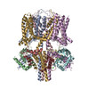
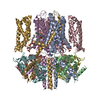
Journal: Proc Natl Acad Sci U S A / Year: 2022 Title: Structural mechanisms for the activation of human cardiac KCNQ1 channel by electro-mechanical coupling enhancers. Authors: Demin Ma / Ling Zhong / Zhenzhen Yan / Jing Yao / Yan Zhang / Fan Ye / Yuan Huang / Dongwu Lai / Wei Yang / Panpan Hou / Jiangtao Guo / Abstract: The cardiac KCNQ1 potassium channel carries the important current and controls the heart rhythm. Hundreds of mutations in KCNQ1 can cause life-threatening cardiac arrhythmia. Although KCNQ1 ...The cardiac KCNQ1 potassium channel carries the important current and controls the heart rhythm. Hundreds of mutations in KCNQ1 can cause life-threatening cardiac arrhythmia. Although KCNQ1 structures have been recently resolved, the structural basis for the dynamic electro-mechanical coupling, also known as the voltage sensor domain-pore domain (VSD-PD) coupling, remains largely unknown. In this study, utilizing two VSD-PD coupling enhancers, namely, the membrane lipid phosphatidylinositol 4,5-bisphosphate (PIP) and a small-molecule ML277, we determined 2.5-3.5 Å resolution cryo-electron microscopy structures of full-length human KCNQ1-calmodulin (CaM) complex in the apo closed, ML277-bound open, and ML277-PIP-bound open states. ML277 binds at the "elbow" pocket above the S4-S5 linker and directly induces an upward movement of the S4-S5 linker and the opening of the activation gate without affecting the C-terminal domain (CTD) of KCNQ1. PIP binds at the cleft between the VSD and the PD and brings a large structural rearrangement of the CTD together with the CaM to activate the PD. These findings not only elucidate the structural basis for the dynamic VSD-PD coupling process during KCNQ1 gating but also pave the way to develop new therapeutics for anti-arrhythmia.
In the structure databanks used in Yorodumi, some data are registered as the other names, "COVID-19 virus" and "2019-nCoV". Here are the details of the virus and the list of structure data.
Jan 31, 2019. EMDB accession codes are about to change! (news from PDBe EMDB page)
EMDB accession codes are about to change! (news from PDBe EMDB page)
The allocation of 4 digits for EMDB accession codes will soon come to an end. Whilst these codes will remain in use, new EMDB accession codes will include an additional digit and will expand incrementally as the available range of codes is exhausted. The current 4-digit format prefixed with “EMD-” (i.e. EMD-XXXX) will advance to a 5-digit format (i.e. EMD-XXXXX), and so on. It is currently estimated that the 4-digit codes will be depleted around Spring 2019, at which point the 5-digit format will come into force.
The EM Navigator/Yorodumi systems omit the EMD- prefix.
Related info.:Q: What is EMD? / ID/Accession-code notation in Yorodumi/EM Navigator
Yorodumi is a browser for structure data from EMDB, PDB, SASBDB, etc.
This page is also the successor to EM Navigator detail page, and also detail information page/front-end page for Omokage search.
The word "yorodu" (or yorozu) is an old Japanese word meaning "ten thousand". "mi" (miru) is to see.
Related info.:EMDB / PDB / SASBDB / Comparison of 3 databanks / Yorodumi Search / Aug 31, 2016. New EM Navigator & Yorodumi / Yorodumi Papers / Jmol/JSmol / Function and homology information / Changes in new EM Navigator and Yorodumi
 Movie
Movie Controller


 Open data
 Basic information
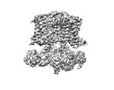
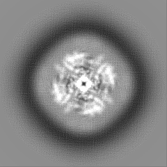
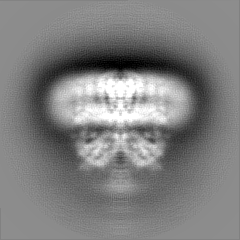
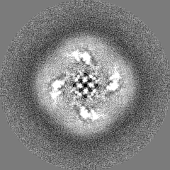
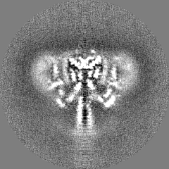
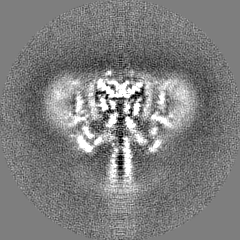
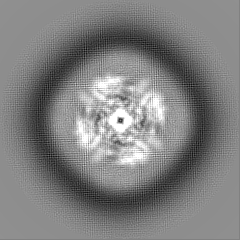
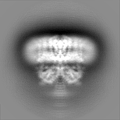
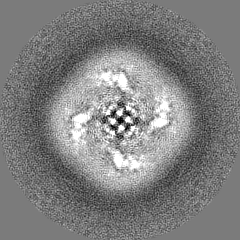
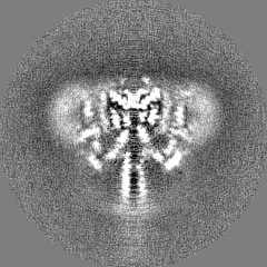
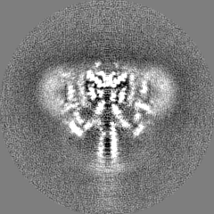
 Map data
 Sample
 Function and homology information
 protein phosphatase 1 binding / positive regulation of potassium ion transmembrane transport / Voltage gated Potassium channels / outward rectifier potassium channel activity / potassium ion homeostasis / ventricular cardiac muscle cell action potential / non-motile cilium assembly / regulation of ventricular cardiac muscle cell membrane repolarization / intestinal absorption /
 Authors
 China, 6 items
  Citation
 Structure visualization
 Downloads & links
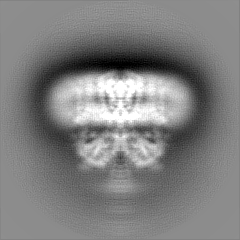
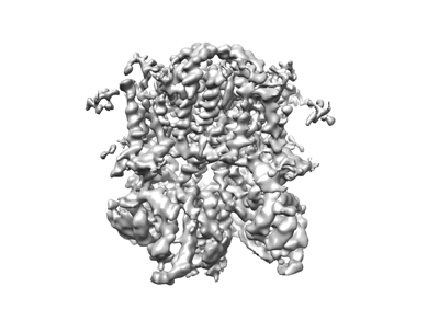 emd_33316.png
 http://ftp.pdbj.org/pub/emdb/structures/EMD-33316


 Z
Z Y
Y X
X


 Sample components
 Processing
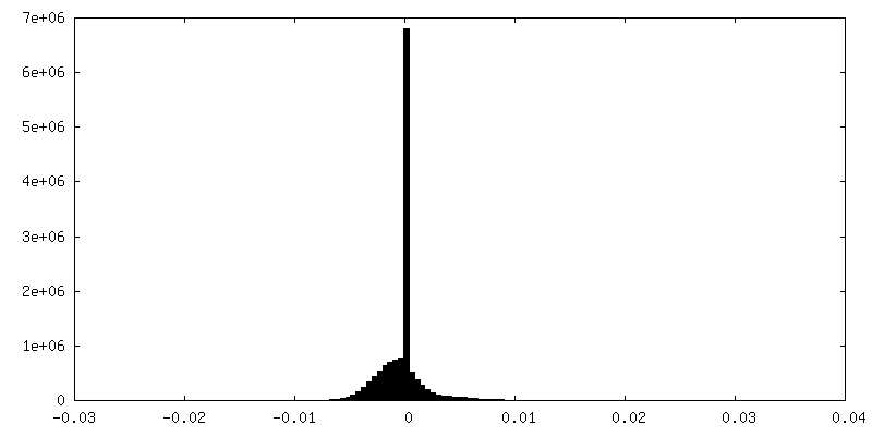
 Electron microscopy