 Movie
Movie Controller

[English] 日本語
 Yorodumi
- PDB-2bk2: The prepore structure of pneumolysin, obtained by fitting the alp... -
+ Open data

- Basic information
| Entry | Database: PDB / ID: 2bk2 | ||||||
|---|---|---|---|---|---|---|---|
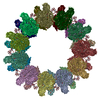
| Title | The prepore structure of pneumolysin, obtained by fitting the alpha carbon trace of perfringolysin O into a cryo-EM map | ||||||
 Components Components | PERFRINGOLYSIN O | ||||||
 Keywords Keywords | TOXIN / CYTOLYSIS / HEMOLYSIS / THIOL-ACTIVATED CYTOLYSIN / CRYOEM / CYTOLYTIC PROTEIN | ||||||
| Function / homology |  Function and homology information Function and homology informationhemolysis in another organism / cholesterol binding / toxin activity / membrane => GO:0016020 / host cell plasma membrane / extracellular region / membrane Similarity search - Function | ||||||
| Biological species |   CLOSTRIDIUM PERFRINGENS (bacteria) CLOSTRIDIUM PERFRINGENS (bacteria) | ||||||
| Method | ELECTRON MICROSCOPY / single particle reconstruction / cryo EM / Resolution: 28 Å | ||||||
| Model type details | CA ATOMS ONLY, CHAIN A | ||||||
 Authors Authors | Tilley, S.J. / Orlova, E.V. / Gilbert, R.J.C. / Andrew, P.W. / Saibil, H.R. | ||||||
 Citation Citation | Journal: Cell / Year: 2005 Title: Structural basis of pore formation by the bacterial toxin pneumolysin. Authors: Sarah J Tilley / Elena V Orlova / Robert J C Gilbert / Peter W Andrew / Helen R Saibil /  Abstract: The bacterial toxin pneumolysin is released as a soluble monomer that kills target cells by assembling into large oligomeric rings and forming pores in cholesterol-containing membranes. Using cryo-EM ...The bacterial toxin pneumolysin is released as a soluble monomer that kills target cells by assembling into large oligomeric rings and forming pores in cholesterol-containing membranes. Using cryo-EM and image processing, we have determined the structures of membrane-surface bound (prepore) and inserted-pore oligomer forms, providing a direct observation of the conformational transition into the pore form of a cholesterol-dependent cytolysin. In the pore structure, the domains of the monomer separate and double over into an arch, forming a wall sealing the bilayer around the pore. This transformation is accomplished by substantial refolding of two of the four protein domains along with deformation of the membrane. Extension of protein density into the bilayer supports earlier predictions that the protein inserts beta hairpins into the membrane. With an oligomer size of up to 44 subunits in the pore, this assembly creates a transmembrane channel 260 A in diameter lined by 176 beta strands. #1: Journal: Cell / Year: 1997Title: Structure of a cholesterol-binding, thiol-activated cytolysin and a model of its membrane form. Authors: J Rossjohn / S C Feil / W J McKinstry / R K Tweten / M W Parker /  Abstract: The mechanisms by which proteins gain entry into membranes is a fundamental problem in biology. Here, we present the first crystal structure of a thiol-activated cytolysin, perfringolysin O, a member ...The mechanisms by which proteins gain entry into membranes is a fundamental problem in biology. Here, we present the first crystal structure of a thiol-activated cytolysin, perfringolysin O, a member of a large family of toxins that kill eukaryotic cells by punching holes in their membranes. The molecule adopts an unusually elongated shape rich in beta sheet. We have used electron microscopy data to construct a detailed model of the membrane channel form of the toxin. The structures reveal a novel mechanism for membrane insertion. Surprisingly, the toxin receptor, cholesterol, appears to play multiple roles: targeting, promotion of oligomerization, triggering a membrane insertion competent form, and stabilizing the membrane pore. | ||||||
| History |
|
- Structure visualization
| Movie |
Movie viewer |
|---|---|
| Structure viewer | Molecule: MolmilJmol/JSmol |
- Downloads & links
-Download
| PDBx/mmCIF format | 2bk2.cif.gz | 30.2 KB | Display | PDBx/mmCIF format |
|---|---|---|---|---|
| PDB format | pdb2bk2.ent.gz | 14.2 KB | Display | PDB format |
| PDBx/mmJSON format | 2bk2.json.gz | Tree view | PDBx/mmJSON format | |
| Others |  Other downloads Other downloads |
-Validation report
| Summary document | 2bk2_validation.pdf.gz | 646 KB | Display | wwPDB validaton report |
|---|---|---|---|---|
| Full document | 2bk2_full_validation.pdf.gz | 645.6 KB | Display | |
| Data in XML | 2bk2_validation.xml.gz | 11.4 KB | Display | |
| Data in CIF | 2bk2_validation.cif.gz | 15.7 KB | Display | |
| Arichive directory | https://data.pdbj.org/pub/pdb/validation_reports/bk/2bk2ftp://data.pdbj.org/pub/pdb/validation_reports/bk/2bk2 | HTTPS FTP |
-Related structure data
| Related structure data |  1106MC 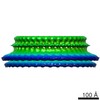 1107C 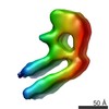 1108C 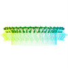 2bk1C C: citing same article ( M: map data used to model this data |
|---|---|
| Similar structure data |


-Links
 PDBj
PDBj- Assembly
| Deposited unit | 
|
|---|---|
| 1 | x 31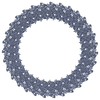
|
| 2 |
|
| 3 | 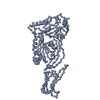
|
| Symmetry | Point symmetry: (Schoenflies symbol: C31 (31 fold cyclic)) |
-Components
| #1: Protein | Mass: 50992.805 Da / Num. of mol.: 1 / Fragment: RESIDUES 36-500 Source method: isolated from a genetically manipulated source Source: (gene. exp.) CLOSTRIDIUM PERFRINGENS (bacteria) / Production host: |
|---|
-Experimental details
-Experiment
| Experiment | Method: ELECTRON MICROSCOPY |
|---|---|
| EM experiment | Aggregation state: PARTICLE / 3D reconstruction method: single particle reconstruction |
- Sample preparation
| Component | Name: PNEUMOLYSIN / Type: COMPLEX Details: THE SAMPLE CONSISTS OF PNEUMOLYSIN IN A MEMBRANE-INSERTED PORE STATE |
|---|---|
| Buffer solution | Name: 8 MM NA2HP04, 1.5MM KH2PO4, 2.5 MM KCL, 0.25 MM NACL / pH: 6.95 Details: 8 MM NA2HP04, 1.5MM KH2PO4, 2.5 MM KCL, 0.25 MM NACL |
| Specimen | Conc.: 0.05 mg/ml / Embedding applied: NO / Shadowing applied: NO / Staining applied: NO / Vitrification applied: YES |
| Specimen support | Details: HOLEY CARBON |
| Vitrification | Instrument: HOMEMADE PLUNGER / Cryogen name: ETHANE / Details: PLUNGED INTO ETHANE |
- Electron microscopy imaging
| Experimental equipment |  Model: Tecnai F20 / Image courtesy: FEI Company |
|---|---|
| Microscopy | Model: FEI TECNAI F20 Details: SAMPLES WERE MAINTAINED AT LIQUID NITROGEN TEMPERATURES IN THE ELECTRON MICROSCOPE. |
| Electron gun | Electron source:  FIELD EMISSION GUN / Accelerating voltage: 200 kV / Illumination mode: FLOOD BEAM FIELD EMISSION GUN / Accelerating voltage: 200 kV / Illumination mode: FLOOD BEAM |
| Electron lens | Mode: BRIGHT FIELD / Nominal magnification: 42000 X / Nominal defocus max: 3200 nm / Nominal defocus min: 1100 nm / Cs: 2 mm |
| Specimen holder | Temperature: 100 K / Tilt angle max: 0 ° / Tilt angle min: 0 ° |
| Image recording | Electron dose: 20 e/Å2 / Film or detector model: KODAK SO-163 FILM |
| Image scans | Num. digital images: 135 |
| Radiation wavelength | Relative weight: 1 |
- Processing
| CTF correction | Details: PHASE FLIPPING | ||||||||||||
|---|---|---|---|---|---|---|---|---|---|---|---|---|---|
| Symmetry | Point symmetry: C31 (31 fold cyclic) | ||||||||||||
| 3D reconstruction | Resolution: 28 Å / Num. of particles: 223 / Nominal pixel size: 3.5 Å Details: RESIDUES 134-142 ARE MISSING FROM THE SEQUENCE. THE 31-MER CAN BE GENERATED BY APPLYING 31-FOLD ROTATIONAL SYMMETRY Symmetry type: POINT | ||||||||||||
| Atomic model building | Protocol: RIGID BODY FIT Details: METHOD--THE CRYSTAL STRUCTURE OF PERFRINOGLYSIN O (1PFO, ROSSJOHN ET AL., 1998, CELL 89, 685)WAS PLACED INTO THE CRYO-EM DENSITY MAP (EMD-1106). THE ALPHA CARBON TRACE OF PERFRINGOLYSIN 0 ...Details: METHOD--THE CRYSTAL STRUCTURE OF PERFRINOGLYSIN O (1PFO, ROSSJOHN ET AL., 1998, CELL 89, 685)WAS PLACED INTO THE CRYO-EM DENSITY MAP (EMD-1106). THE ALPHA CARBON TRACE OF PERFRINGOLYSIN 0 WAS MANUALLY POSITIONED INTO THE CRYO-EM DENSITY CORRESPONDING TO THE POSITION OF ONE SUBUNIT. THE BEST FIT WAS OBTAINED BY SEPARATING THE MONOMER INTO TWO RIGID BODIES- DOMAINS 1-3 (36-390) AND DOMAIN 4 (391-500). THE COMPLETE OLIGOMER (31-MER) WAS GENERATED AND CHECKED FOR CLOSE CONTACTS BOTH BY EYE AND USING THE CCP4 PROGRAM CONTACT. | ||||||||||||
| Atomic model building | PDB-ID: 1PFO Accession code: 1PFO / Source name: PDB / Type: experimental model | ||||||||||||
| Refinement | Highest resolution: 28 Å | ||||||||||||
| Refinement step | Cycle: LAST / Highest resolution: 28 Å
|
