 Movie
Movie Controller


+ Open data

- Basic information
| Entry | Database: EMDB / ID: EMD-2755 | |||||||||
|---|---|---|---|---|---|---|---|---|---|---|
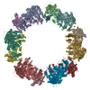
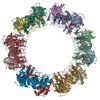
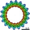
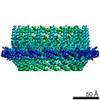
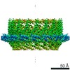
| Title | Cryo-electron microscopy of TibC dodecamer | |||||||||
 Map data Map data | Reconstruction of TibC dodecamer | |||||||||
 Sample Sample |
| |||||||||
 Keywords Keywords | Bacterial autotransporters / Glycosyltransferase / Bacterial pathogenesis / Cryo-EM / Enzyme complex | |||||||||
| Biological species |  | |||||||||
| Method | single particle reconstruction / cryo EM / Resolution: 11.5 Å | |||||||||
 Authors Authors | Yao Q / Lu QH / Wan XB / Song F / Xu Y / Zamyatina A / Huang N / Zhu P / Shao F | |||||||||
 Citation Citation | Journal: Elife / Year: 2014 Title: A structural mechanism for bacterial autotransporter glycosylation by a dodecameric heptosyltransferase family. Authors: Qing Yao / Qiuhe Lu / Xiaobo Wan / Feng Song / Yue Xu / Mo Hu / Alla Zamyatina / Xiaoyun Liu / Niu Huang / Ping Zhu / Feng Shao /   Abstract: A large group of bacterial virulence autotransporters including AIDA-I from diffusely adhering E. coli (DAEC) and TibA from enterotoxigenic E. coli (ETEC) require hyperglycosylation for functioning. ...A large group of bacterial virulence autotransporters including AIDA-I from diffusely adhering E. coli (DAEC) and TibA from enterotoxigenic E. coli (ETEC) require hyperglycosylation for functioning. Here we demonstrate that TibC from ETEC harbors a heptosyltransferase activity on TibA and AIDA-I, defining a large family of bacterial autotransporter heptosyltransferases (BAHTs). The crystal structure of TibC reveals a characteristic ring-shape dodecamer. The protomer features an N-terminal β-barrel, a catalytic domain, a β-hairpin thumb, and a unique iron-finger motif. The iron-finger motif contributes to back-to-back dimerization; six dimers form the ring through β-hairpin thumb-mediated hand-in-hand contact. The structure of ADP-D-glycero-β-D-manno-heptose (ADP-D,D-heptose)-bound TibC reveals a sugar transfer mechanism and also the ligand stereoselectivity determinant. Electron-cryomicroscopy analyses uncover a TibC-TibA dodecamer/hexamer assembly with two enzyme molecules binding to one TibA substrate. The complex structure also highlights a high efficient hyperglycosylation of six autotransporter substrates simultaneously by the dodecamer enzyme complex. | |||||||||
| History |
|
- Structure visualization
| Movie |
 Movie viewer Movie viewer |
|---|---|
| Structure viewer | EM map: SurfViewMolmilJmol/JSmol |
| Supplemental images |


- Downloads & links
-EMDB archive
| Map data | emd_2755.map.gz | 45.9 MB | EMDB map data format | |
|---|---|---|---|---|
| Header (meta data) | emd-2755-v30.xmlemd-2755.xml | 9.1 KB 9.1 KB | Display Display | EMDB header |
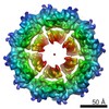
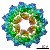
| Images | 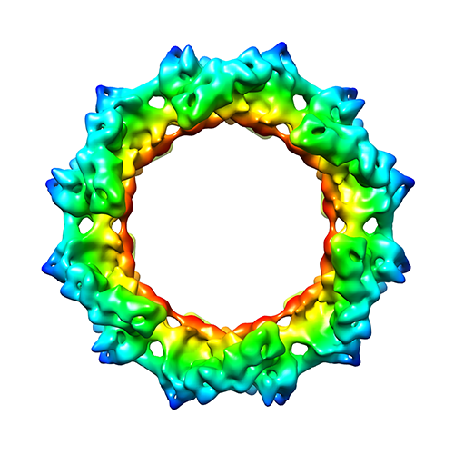 EMD-2755.png EMD-2755.png emd_2755.png emd_2755.png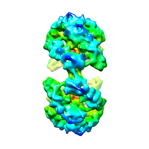 emd_2755_1.png emd_2755_1.png | 151 KB 151 KB 123.8 KB | ||
| Archive directory |  http://ftp.pdbj.org/pub/emdb/structures/EMD-2755ftp://ftp.pdbj.org/pub/emdb/structures/EMD-2755 http://ftp.pdbj.org/pub/emdb/structures/EMD-2755ftp://ftp.pdbj.org/pub/emdb/structures/EMD-2755 | HTTPS FTP |
-Validation report
| Summary document | emd_2755_validation.pdf.gz | 218.3 KB | Display | EMDB validaton report |
|---|---|---|---|---|
| Full document | emd_2755_full_validation.pdf.gz | 217.4 KB | Display | |
| Data in XML | emd_2755_validation.xml.gz | 6.5 KB | Display | |
| Arichive directory | https://ftp.pdbj.org/pub/emdb/validation_reports/EMD-2755ftp://ftp.pdbj.org/pub/emdb/validation_reports/EMD-2755 | HTTPS FTP |
-Related structure data


-Links
| EMDB pages | EMDB (EBI/PDBe) / EMDataResource |
|---|
-Map
| File | Download / File: emd_2755.map.gz / Format: CCP4 / Size: 51.5 MB / Type: IMAGE STORED AS FLOATING POINT NUMBER (4 BYTES) | ||||||||||||||||||||||||||||||||||||||||||||||||||||||||||||||||||||
|---|---|---|---|---|---|---|---|---|---|---|---|---|---|---|---|---|---|---|---|---|---|---|---|---|---|---|---|---|---|---|---|---|---|---|---|---|---|---|---|---|---|---|---|---|---|---|---|---|---|---|---|---|---|---|---|---|---|---|---|---|---|---|---|---|---|---|---|---|---|
| Annotation | Reconstruction of TibC dodecamer | ||||||||||||||||||||||||||||||||||||||||||||||||||||||||||||||||||||
| Voxel size | X=Y=Z: 1.196 Å | ||||||||||||||||||||||||||||||||||||||||||||||||||||||||||||||||||||
| Density |
| ||||||||||||||||||||||||||||||||||||||||||||||||||||||||||||||||||||
| Symmetry | Space group: 1 | ||||||||||||||||||||||||||||||||||||||||||||||||||||||||||||||||||||
| Details | EMDB XML:
CCP4 map header:
| ||||||||||||||||||||||||||||||||||||||||||||||||||||||||||||||||||||
-Supplemental data
- Sample components
-Entire : Complex of TibC
| Entire | Name: Complex of TibC |
|---|---|
| Components |
|
-Supramolecule #1000: Complex of TibC
| Supramolecule | Name: Complex of TibC / type: sample / ID: 1000 / Oligomeric state: TibC forms a dodecamer of six dimers / Number unique components: 1 |
|---|---|
| Molecular weight | Theoretical: 555 KDa |
-Macromolecule #1: TibC
| Macromolecule | Name: TibC / type: protein_or_peptide / ID: 1 Details: Ferric ions were attached to specific cysteine residues Number of copies: 12 / Oligomeric state: Dodecamer / Recombinant expression: Yes |
|---|---|
| Source (natural) | Organism: |
| Molecular weight | Theoretical: 46 KDa |
| Recombinant expression | Organism: |
-Experimental details
-Structure determination
| Method | cryo EM |
|---|---|
 Processing Processing | single particle reconstruction |
| Aggregation state | particle |
-Sample preparation
| Concentration | 1 mg/mL |
|---|---|
| Buffer | pH: 7.6 / Details: 10mM Tris-HCl, 100mM NaCl, 2mM DTT |
| Grid | Details: Quantifoil R2.1, 300 mesh |
| Vitrification | Cryogen name: ETHANE / Chamber humidity: 100 % / Instrument: FEI VITROBOT MARK IV Method: 10 ug/ml bacitracin (Sigma) was added to the purified protein to obtain monodispersed particles and make the orientation distribution more anisotropic. Blot for 4 sec using blotting force 2 before plunging. |
- Electron microscopy
| Microscope | FEI TITAN KRIOS |
|---|---|
| Alignment procedure | Legacy - Astigmatism: Objective lens astigmatism was corrected at 155,000 times magnification |
| Date | Oct 11, 2012 |
| Image recording | Category: CCD / Film or detector model: GATAN ULTRASCAN 4000 (4k x 4k) / Number real images: 3000 / Average electron dose: 18 e/Å2 |
| Electron beam | Acceleration voltage: 300 kV / Electron source:  FIELD EMISSION GUN FIELD EMISSION GUN |
| Electron optics | Illumination mode: FLOOD BEAM / Imaging mode: BRIGHT FIELD / Cs: 2.7 mm / Nominal magnification: 75000 |
| Sample stage | Specimen holder model: FEI TITAN KRIOS AUTOGRID HOLDER |
| Experimental equipment |  Model: Titan Krios / Image courtesy: FEI Company |
-Image processing
| CTF correction | Details: CTF correction of each particle |
|---|---|
| Final reconstruction | Applied symmetry - Point group: C6 (6 fold cyclic) / Algorithm: OTHER / Resolution.type: BY AUTHOR / Resolution: 11.5 Å / Resolution method: OTHER / Software - Name: EMAN2 / Number images used: 8546 |
