 Movie
Movie Controller

[English] 日本語
 Yorodumi
- EMDB-22077: Cryo-EM Structure of CagX and CagY within the dCag3 Helicobacter ... -
+ Open data

- Basic information
| Entry | Database: EMDB / ID: EMD-22077 | ||||||||||||||||||||||||
|---|---|---|---|---|---|---|---|---|---|---|---|---|---|---|---|---|---|---|---|---|---|---|---|---|---|
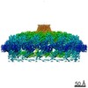
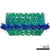
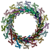
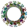
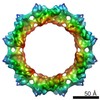
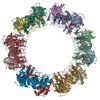
| Title | Cryo-EM Structure of CagX and CagY within the dCag3 Helicobacter pylori PR | ||||||||||||||||||||||||
 Map data Map data | Helicobacter pylori T4SS dCag3 PR | ||||||||||||||||||||||||
 Sample Sample |
| ||||||||||||||||||||||||
| Function / homology |  Function and homology information Function and homology information | ||||||||||||||||||||||||
| Biological species |    Helicobacter pylori (bacteria) / Helicobacter pylori (strain ATCC 700392 / 26695) (bacteria) Helicobacter pylori (bacteria) / Helicobacter pylori (strain ATCC 700392 / 26695) (bacteria) | ||||||||||||||||||||||||
| Method | single particle reconstruction / cryo EM / Resolution: 3.0 Å | ||||||||||||||||||||||||
 Authors Authors | Sheedlo MJ / Chung JM / Sawhney N / Durie CL / Cover TL / Ohi MD / Lacy DB | ||||||||||||||||||||||||
| Funding support |  United States, 7 items United States, 7 items
| ||||||||||||||||||||||||
 Citation Citation | Journal: Elife / Year: 2020 Title: Cryo-EM reveals species-specific components within the Cag type IV secretion system core complex. Authors: Michael J Sheedlo / Jeong Min Chung / Neha Sawhney / Clarissa L Durie / Timothy L Cover / Melanie D Ohi / D Borden Lacy / Abstract: The pathogenesis of -associated gastric cancer is dependent on delivery of CagA into host cells through a type IV secretion system (T4SS). The Cag T4SS includes a large membrane-spanning core ...The pathogenesis of -associated gastric cancer is dependent on delivery of CagA into host cells through a type IV secretion system (T4SS). The Cag T4SS includes a large membrane-spanning core complex containing five proteins, organized into an outer membrane cap (OMC), a periplasmic ring (PR) and a stalk. Here, we report cryo-EM reconstructions of a core complex lacking Cag3 and an improved map of the wild-type complex. We define the structures of two unique species-specific components (Cag3 and CagM) and show that Cag3 is structurally similar to CagT. Unexpectedly, components of the OMC are organized in a 1:1:2:2:5 molar ratio (CagY:CagX:CagT:CagM:Cag3). CagX and CagY are components of both the OMC and the PR and bridge the symmetry mismatch between these regions. These results reveal that assembly of the T4SS core complex is dependent on incorporation of interwoven species-specific components. | ||||||||||||||||||||||||
| History |
|
- Structure visualization
| Movie |
Movie viewer |
|---|---|
| Structure viewer | EM map: SurfViewMolmilJmol/JSmol |
| Supplemental images |

- Downloads & links
-EMDB archive
| Map data | emd_22077.map.gz | 473.4 MB | EMDB map data format | |
|---|---|---|---|---|
| Header (meta data) | emd-22077-v30.xmlemd-22077.xml | 13.6 KB 13.6 KB | Display Display | EMDB header |
| FSC (resolution estimation) | emd_22077_fsc.xml | 18 KB | Display | FSC data file |
| Images | 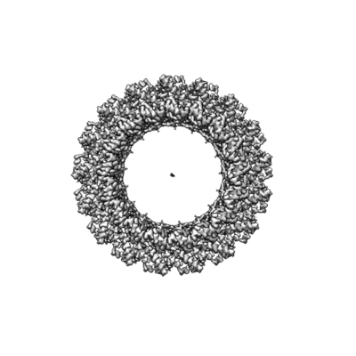 emd_22077.png emd_22077.png | 100.3 KB | ||
| Archive directory |  http://ftp.pdbj.org/pub/emdb/structures/EMD-22077ftp://ftp.pdbj.org/pub/emdb/structures/EMD-22077 http://ftp.pdbj.org/pub/emdb/structures/EMD-22077ftp://ftp.pdbj.org/pub/emdb/structures/EMD-22077 | HTTPS FTP |
-Related structure data
| Related structure data |  6x6lMC  6x6jC  6x6kC  6x6sC M: atomic model generated by this map C: citing same article ( |
|---|---|
| Similar structure data |


-Links
| EMDB pages | EMDB (EBI/PDBe) / EMDataResource |
|---|
-Map
| File | Download / File: emd_22077.map.gz / Format: CCP4 / Size: 506 MB / Type: IMAGE STORED AS FLOATING POINT NUMBER (4 BYTES) | ||||||||||||||||||||||||||||||||||||||||||||||||||||||||||||||||||||
|---|---|---|---|---|---|---|---|---|---|---|---|---|---|---|---|---|---|---|---|---|---|---|---|---|---|---|---|---|---|---|---|---|---|---|---|---|---|---|---|---|---|---|---|---|---|---|---|---|---|---|---|---|---|---|---|---|---|---|---|---|---|---|---|---|---|---|---|---|---|
| Annotation | Helicobacter pylori T4SS dCag3 PR | ||||||||||||||||||||||||||||||||||||||||||||||||||||||||||||||||||||
| Voxel size | X=Y=Z: 1 Å | ||||||||||||||||||||||||||||||||||||||||||||||||||||||||||||||||||||
| Density |
| ||||||||||||||||||||||||||||||||||||||||||||||||||||||||||||||||||||
| Symmetry | Space group: 1 | ||||||||||||||||||||||||||||||||||||||||||||||||||||||||||||||||||||
| Details | EMDB XML:
CCP4 map header:
| ||||||||||||||||||||||||||||||||||||||||||||||||||||||||||||||||||||
-Supplemental data
- Sample components
-Entire : Helicobacter pylori dCag3 Cag T4SS PR
| Entire | Name: Helicobacter pylori dCag3 Cag T4SS PR |
|---|---|
| Components |
|
-Supramolecule #1: Helicobacter pylori dCag3 Cag T4SS PR
| Supramolecule | Name: Helicobacter pylori dCag3 Cag T4SS PR / type: complex / ID: 1 / Parent: 0 / Macromolecule list: all |
|---|---|
| Source (natural) | Organism: Helicobacter pylori (bacteria) |
-Macromolecule #1: Cag pathogenicity island protein (Cag8)
| Macromolecule | Name: Cag pathogenicity island protein (Cag8) / type: protein_or_peptide / ID: 1 / Number of copies: 17 / Enantiomer: LEVO |
|---|---|
| Source (natural) | Organism: Helicobacter pylori (strain ATCC 700392 / 26695) (bacteria) Strain: ATCC 700392 / 26695 |
| Molecular weight | Theoretical: 60.607324 KDa |
| Sequence | String: MGQAFFKKIV GCFCLGYLFL SSAIEAAALD IKNFNRGRVK VVNKKIAYLG DEKPITIWTS LDNVTVIQLE KDETISYITT GFNKGWSIV PNSNHIFIQP KSVKSNLMFE KEAVNFALMT RDYQEFLKTK KLIVDAPDPK ELEEQKKALE KEKEAKEQAQ K AQKDKREK ...String: MGQAFFKKIV GCFCLGYLFL SSAIEAAALD IKNFNRGRVK VVNKKIAYLG DEKPITIWTS LDNVTVIQLE KDETISYITT GFNKGWSIV PNSNHIFIQP KSVKSNLMFE KEAVNFALMT RDYQEFLKTK KLIVDAPDPK ELEEQKKALE KEKEAKEQAQ K AQKDKREK RKEERAKNRA NLENLTNAMS NPQNLSNNKN LSEFIKQQRE NELDQMERLE DMQEQAQANA LKQIEELNKK QA EETIKQR AKDKINIKTD KPQKSPEDNS IELSPSDSAW RTNLVVRTNK ALYQFILRIA QKDNFASAYL TVKLEYPQRH EVS SVIEEL KKREEAKRQK ELIKQENLNT TAYINRVMMA SNEQIINKEK IREEKQKIIL DQAKALETQY VHNALKRNPV PRNY NYYQA PEKRSKHIMP SEIFDDGTFT YFGFKNITLQ PAIFVVQPDG KLSMTDAAID PNMTNSGLRW YRVNEIAEKF KLIKD KALV TVINKGYGKN PLTKNYNIKN YGELERVIKK LPEVRDK |
-Macromolecule #2: Cag pathogenicity island protein (Cag7)
| Macromolecule | Name: Cag pathogenicity island protein (Cag7) / type: protein_or_peptide / ID: 2 / Number of copies: 17 / Enantiomer: LEVO |
|---|---|
| Source (natural) | Organism: Helicobacter pylori (strain ATCC 700392 / 26695) (bacteria) Strain: ATCC 700392 / 26695 |
| Molecular weight | Theoretical: 219.748641 KDa |
| Sequence | String: MNEENDKLET SKKAQQDSPQ DLSNEEATEA NHFENLLKES KESSDHHLDN PTETQTHFDG DKSEETQTQM DSEGNETSES SNGSLADKL FKKARKLVDN KKPFTQQKNL DEETQELNEE DDQENNEYQE ETQTDLIDDE TSKKTQQHSP QDLSNEEATE A NHFENLLK ...String: MNEENDKLET SKKAQQDSPQ DLSNEEATEA NHFENLLKES KESSDHHLDN PTETQTHFDG DKSEETQTQM DSEGNETSES SNGSLADKL FKKARKLVDN KKPFTQQKNL DEETQELNEE DDQENNEYQE ETQTDLIDDE TSKKTQQHSP QDLSNEEATE A NHFENLLK ESKESSDHHL DNPTETQTNF DGDKSEETQT QMDSEGNETS ESSNGSLADK LFKKARKLVD NKKPFTQQKN LD EETQELN EEDDQENNEY QEETQTDLID DETSKKTQQH SPQDLSNEEA TEANHFENLL KESKESSDHH LDNPTETQTN FDG DKSEEI TDDSNDQEII KGSKKKYIIG GIVVAVLIVI ILFSRSIFHY FMPLEDKSSR FSKDRNLYVN DEIQIRQEYN RLLK ERNEK GNMIDKNLFF NDDPNRTLYN YLNIAEIEDK NPLRAFYECI SNGGNYEECL KLIKDKKLQD QMKKTLEAYN DCIKN AKTE EERIKCLDLI KDENLKKSLL NQQKVQVALD CLKNAKTDEE RNECLKLIND PEIREKFRKE LELQKELQEY KDCIKN AKT EAEKNKCLKG LSKEAIERLK QQALDCLKNA KTDEERNECL KNIPQDLQKE LLADMSVKAY KDCVSKARNE KEKQECE KL LTPEARKKLE QQVLDCLKNA KTDEERKKCL KDLPKDLQSD ILAKESLKAY KDCVSQAKTE AEKKECEKLL TPEAKKLL E EEAKESVKAY LDCVSQAKTE AEKKECEKLL TPEAKKKLEE AKKSVKAYLD CVSRARNEKE KKECEKLLTP EAKKLLEQQ ALDCLKNAKT DKERKKCLKD LPKDLQKKVL AKESVKAYLD CVSQAKTEAE KKECEKLLTP EARKLLEEAK KSVKAYLDCV SQAKTEAEK KECEKLLTPE ARKLLEE(UNK)AK ESVKAYLDCV SQAKNEAEKK ECEKLLTLES KKKLEEAKKS VKAYLDC VS QAKTEAEKKE CEKLLTPEAK KLLEQQALDC LKNAKTEADK KRCVKDLPKD LQKKVLAKES LKAYKDCVSK ARNEKEKK E CEKLLTPEAK KLLEEAKKSV KAYLDCVSQA KTEAEKKECE KLLTPEARKL LEEAKESVKA YKDCVSKARN EKEKKECEK LLTPEAKKLL EQQVLDCLKN AKTEADKKRC VKDLPKDLQK KVLAKESVKA YLDCVSRARN EKEKKECEKL LTPEAKKLLE EAKESLKAY KDCLSQARNE EERRACEKLL TPEARKLLEQ EVKKSIKAYL DCVSRARNEK EKKECEKLLT PEARKFLAKQ V LNCLEKAG NEEERKACLK NLPKDLQENI LAKESLKAYK DCLSQARNEE ERRACEKLLT PEARKLLEQE VKKSVKAYLD CV SRARNEK EKKECEKLLT PEARKFLAKE LQQKDKAIKD CLKNADPNDR AAIMKCLDGL SDEEKLKYLQ EAREKAVADC LAM AKTDEE KRKCQNLYSD LIQEIQNKRT QNKQNQLSKT ERLHQASECL DNLDDPTDQE AIEQCLEGLS DSERALILGI KRQA DEVDL IYSDLRNRKT FDNMAAKGYP LLPMDFKNGG DIATINATNV DADKIASDNP IYASIEPDIA KQYETEKTIK DKNLE AKLA KALGGNKKDD DKEKSKKSTA EAKAENNKID KDVAETAKNI SEIALKNKKE KSGEFVDENG NPIDDKKKAE KQDETS PVK QAFIGKSDPT FVLAQYTPIE ITLTSKVDAT LTGIVSGVVA KDVWNMNGTM ILLDKGTKVY GNYQSVKGGT PIMTRLM IV FTKAITPDGV IIPLANAQAA GMLGEAGVDG YVNNHFMKRI GFAVIASVVN SFLQTAPIIA LDKLIGLGKG RSERTPEF N YALGQAINGS MQSSAQMSNQ ILGQLMNIPP SFYKNEGDSI KILTMDDIDF SGVYDVKITN KSVVDEIIKQ STKTLSREH EEITTSPKGG N |
-Experimental details
-Structure determination
| Method | cryo EM |
|---|---|
 Processing Processing | single particle reconstruction |
| Aggregation state | particle |
-Sample preparation
| Buffer | pH: 7 |
|---|---|
| Vitrification | Cryogen name: ETHANE |
- Electron microscopy
| Microscope | FEI TITAN KRIOS |
|---|---|
| Electron beam | Acceleration voltage: 300 kV / Electron source: FIELD EMISSION GUN |
| Electron optics | Illumination mode: FLOOD BEAM / Imaging mode: BRIGHT FIELDBright-field microscopy |
| Image recording | Film or detector model: GATAN K2 SUMMIT (4k x 4k) / Average electron dose: 59.7 e/Å2 |
| Experimental equipment |  Model: Titan Krios / Image courtesy: FEI Company |
-Image processing
| Initial angle assignment | Type: MAXIMUM LIKELIHOOD |
|---|---|
| Final angle assignment | Type: MAXIMUM LIKELIHOOD |
| Final reconstruction | Resolution.type: BY AUTHOR / Resolution: 3.0 Å / Resolution method: FSC 0.143 CUT-OFF / Number images used: 10477 |
| FSC plot (resolution estimation) |  |
