Movie
Movie Controller

[English] 日本語
 Yorodumi
- EMDB-26772: Cryogenic electron microscopy 3D map of F-actin bound by human di... -
+ Open data

- Basic information
| Entry |  | |||||||||
|---|---|---|---|---|---|---|---|---|---|---|
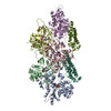
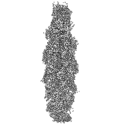
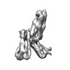
| Title | Cryogenic electron microscopy 3D map of F-actin bound by human dimeric alpha-catenin | |||||||||
 Map data Map data | Cryoem map | |||||||||
 Sample Sample |
| |||||||||
| Function / homology |  Function and homology information Function and homology informationnegative regulation of integrin-mediated signaling pathway / CDH11 homotypic and heterotypic interactions / Regulation of CDH19 Expression and Function / Regulation of CDH11 function / gamma-catenin binding / epithelial cell-cell adhesion /  zonula adherens / gap junction assembly / cellular response to indole-3-methanol / vinculin binding ...negative regulation of integrin-mediated signaling pathway / CDH11 homotypic and heterotypic interactions / Regulation of CDH19 Expression and Function / Regulation of CDH11 function / gamma-catenin binding / epithelial cell-cell adhesion / zonula adherens / gap junction assembly / cellular response to indole-3-methanol / vinculin binding / flotillin complex / negative regulation of cell motility / apical junction assembly / positive regulation of extrinsic apoptotic signaling pathway in absence of ligand / positive regulation of smoothened signaling pathway / Adherens junctions interactions / catenin complex / negative regulation of protein localization to nucleus / axon regeneration / cytoskeletal motor activator activity / negative regulation of neuroblast proliferation / smoothened signaling pathway / establishment or maintenance of cell polarity / Myogenesis / tropomyosin binding / mesenchyme migration / myosin heavy chain binding / troponin I binding / odontogenesis of dentin-containing tooth / filamentous actin / actin filament bundle / skeletal muscle thin filament assembly / striated muscle thin filament / actin filament bundle assembly / skeletal muscle myofibril / intercalated disc / actin monomer binding / negative regulation of extrinsic apoptotic signaling pathway in absence of ligand / neuroblast proliferation / RHO GTPases activate IQGAPs / skeletal muscle fiber development / stress fiber / ovarian follicle development / titin binding / extrinsic apoptotic signaling pathway in absence of ligand / actin filament polymerization / acrosomal vesicle / VEGFR2 mediated vascular permeability / filopodium / integrin-mediated signaling pathway / actin filament / adherens junction / Hydrolases; Acting on acid anhydrides; Acting on acid anhydrides to facilitate cellular and subcellular movement / protein localization / beta-catenin binding / cell-cell adhesion / response to estrogen / calcium-dependent protein binding / male gonad development / actin filament binding / cell-cell junction / cell migration / actin cytoskeleton / cell junction / lamellipodium / cell body / hydrolase activity / cell adhesion / cadherin binding / protein domain specific binding / intracellular membrane-bounded organelle / focal adhesion / calcium ion binding / positive regulation of gene expression / structural molecule activity / magnesium ion binding / RNA binding / ATP binding / identical protein binding / plasma membrane / cytosol / cytoplasm zonula adherens / gap junction assembly / cellular response to indole-3-methanol / vinculin binding ...negative regulation of integrin-mediated signaling pathway / CDH11 homotypic and heterotypic interactions / Regulation of CDH19 Expression and Function / Regulation of CDH11 function / gamma-catenin binding / epithelial cell-cell adhesion / zonula adherens / gap junction assembly / cellular response to indole-3-methanol / vinculin binding / flotillin complex / negative regulation of cell motility / apical junction assembly / positive regulation of extrinsic apoptotic signaling pathway in absence of ligand / positive regulation of smoothened signaling pathway / Adherens junctions interactions / catenin complex / negative regulation of protein localization to nucleus / axon regeneration / cytoskeletal motor activator activity / negative regulation of neuroblast proliferation / smoothened signaling pathway / establishment or maintenance of cell polarity / Myogenesis / tropomyosin binding / mesenchyme migration / myosin heavy chain binding / troponin I binding / odontogenesis of dentin-containing tooth / filamentous actin / actin filament bundle / skeletal muscle thin filament assembly / striated muscle thin filament / actin filament bundle assembly / skeletal muscle myofibril / intercalated disc / actin monomer binding / negative regulation of extrinsic apoptotic signaling pathway in absence of ligand / neuroblast proliferation / RHO GTPases activate IQGAPs / skeletal muscle fiber development / stress fiber / ovarian follicle development / titin binding / extrinsic apoptotic signaling pathway in absence of ligand / actin filament polymerization / acrosomal vesicle / VEGFR2 mediated vascular permeability / filopodium / integrin-mediated signaling pathway / actin filament / adherens junction / Hydrolases; Acting on acid anhydrides; Acting on acid anhydrides to facilitate cellular and subcellular movement / protein localization / beta-catenin binding / cell-cell adhesion / response to estrogen / calcium-dependent protein binding / male gonad development / actin filament binding / cell-cell junction / cell migration / actin cytoskeleton / cell junction / lamellipodium / cell body / hydrolase activity / cell adhesion / cadherin binding / protein domain specific binding / intracellular membrane-bounded organelle / focal adhesion / calcium ion binding / positive regulation of gene expression / structural molecule activity / magnesium ion binding / RNA binding / ATP binding / identical protein binding / plasma membrane / cytosol / cytoplasmSimilarity search - Function | |||||||||
| Biological species |  Homo sapiens (human) / Homo sapiens (human) /  Oryctolagus cuniculus (rabbit) Oryctolagus cuniculus (rabbit) | |||||||||
| Method | helical reconstruction / cryo EM / Resolution: 2.77 Å | |||||||||
 Authors Authors | Rangarajan ES / Smith EW / Izard T | |||||||||
| Funding support |  United States, 1 items United States, 1 items
| |||||||||
 Citation Citation | Journal: Commun Biol / Year: 2023 Title: Distinct inter-domain interactions of dimeric versus monomeric α-catenin link cell junctions to filaments. Authors: Erumbi S Rangarajan / Emmanuel W Smith / Tina Izard / Abstract: Attachment between cells is crucial for almost all aspects of the life of cells. These inter-cell adhesions are mediated by the binding of transmembrane cadherin receptors of one cell to cadherins of ...Attachment between cells is crucial for almost all aspects of the life of cells. These inter-cell adhesions are mediated by the binding of transmembrane cadherin receptors of one cell to cadherins of a neighboring cell. Inside the cell, cadherin binds β-catenin, which interacts with α-catenin. The transitioning of cells between migration and adhesion is modulated by α-catenin, which links cell junctions and the plasma membrane to the actin cytoskeleton. At cell junctions, a single β-catenin/α-catenin heterodimer slips along filamentous actin in the direction of cytoskeletal tension which unfolds clustered heterodimers to form catch bonds with F-actin. Outside cell junctions, α-catenin dimerizes and links the plasma membrane to F-actin. Under cytoskeletal tension, α-catenin unfolds and forms an asymmetric catch bond with F-actin. To understand the mechanism of this important α-catenin function, we determined the 2.7 Å cryogenic electron microscopy (cryoEM) structures of filamentous actin alone and bound to human dimeric α-catenin. Our structures provide mechanistic insights into the role of the α-catenin interdomain interactions in directing α-catenin function and suggest a bivalent mechanism. Further, our cryoEM structure of human monomeric α-catenin provides mechanistic insights into α-catenin autoinhibition. Collectively, our structures capture the initial α-catenin interaction with F-actin before the sensing of force, which is a crucial event in cell adhesion and human disease. | |||||||||
| History |
|
- Structure visualization
| Supplemental images |
|---|
- Downloads & links
-EMDB archive
| Map data | emd_26772.map.gz | 167.9 MB | EMDB map data format | |
|---|---|---|---|---|
| Header (meta data) | emd-26772-v30.xmlemd-26772.xml | 19.6 KB 19.6 KB | Display Display | EMDB header |
| FSC (resolution estimation) | emd_26772_fsc.xml | 16.4 KB | Display | FSC data file |
| Images |  emd_26772.png emd_26772.png | 96 KB | ||
| Others | emd_26772_half_map_1.map.gzemd_26772_half_map_2.map.gz | 165.4 MB 165.4 MB | ||
| Archive directory |  http://ftp.pdbj.org/pub/emdb/structures/EMD-26772ftp://ftp.pdbj.org/pub/emdb/structures/EMD-26772 http://ftp.pdbj.org/pub/emdb/structures/EMD-26772ftp://ftp.pdbj.org/pub/emdb/structures/EMD-26772 | HTTPS FTP |
-Related structure data
| Related structure data | 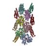 7utjMC  7uxfC M: atomic model generated by this map C: citing same article ( |
|---|---|
| Similar structure data |


-Links
| EMDB pages | EMDB (EBI/PDBe) / EMDataResource |
|---|---|
| Related items in Molecule of the Month |


-Map
| File | Download / File: emd_26772.map.gz / Format: CCP4 / Size: 178 MB / Type: IMAGE STORED AS FLOATING POINT NUMBER (4 BYTES) | ||||||||||||||||||||
|---|---|---|---|---|---|---|---|---|---|---|---|---|---|---|---|---|---|---|---|---|---|
| Annotation | Cryoem map | ||||||||||||||||||||
| Voxel size | X=Y=Z: 1.087 Å | ||||||||||||||||||||
| Density |
| ||||||||||||||||||||
| Symmetry | Space group: 1 | ||||||||||||||||||||
| Details | EMDB XML:
|
-Supplemental data
-Half map: Half map 1
| File | emd_26772_half_map_1.map | ||||||||||||
|---|---|---|---|---|---|---|---|---|---|---|---|---|---|
| Annotation | Half map 1 | ||||||||||||
| Projections & Slices |
| ||||||||||||
| Density Histograms |
 Z
Z Y
Y X
X


-Half map: Half map 2
| File | emd_26772_half_map_2.map | ||||||||||||
|---|---|---|---|---|---|---|---|---|---|---|---|---|---|
| Annotation | Half map 2 | ||||||||||||
| Projections & Slices |
| ||||||||||||
| Density Histograms |


- Sample components
-Entire : dimeric alpha-catenin (residues 22-906) complexed with F-actin
| Entire | Name: dimeric alpha-catenin (residues 22-906) complexed with F-actin |
|---|---|
| Components |
|
-Supramolecule #1: dimeric alpha-catenin (residues 22-906) complexed with F-actin
| Supramolecule | Name: dimeric alpha-catenin (residues 22-906) complexed with F-actin type: complex / ID: 1 / Chimera: Yes / Parent: 0 / Macromolecule list: #1-#2 |
|---|---|
| Source (natural) | Organism: Homo sapiens (human) |
-Macromolecule #1: Actin, alpha skeletal muscle
| Macromolecule | Name: Actin, alpha skeletal muscle / type: protein_or_peptide / ID: 1 / Number of copies: 6 / Enantiomer: LEVO |
|---|---|
| Source (natural) | Organism: Oryctolagus cuniculus (rabbit) |
| Molecular weight | Theoretical: 42.109973 KDa |
| Recombinant expression | Organism:  Escherichia coli (E. coli) Escherichia coli (E. coli) |
| Sequence | String: MCDEDETTAL VCDNGSGLVK AGFAGDDAPR AVFPSIVGRP RHQGVMVGMG QKDSYVGDEA QSKRGILTLK YPIE(HIC)G IIT NWDDMEKIWH HTFYNELRVA PEEHPTLLTE APLNPKANRE KMTQIMFETF NVPAMYVAIQ AVLSLYASGR TTGIVLD SG DGVTHNVPIY ...String: MCDEDETTAL VCDNGSGLVK AGFAGDDAPR AVFPSIVGRP RHQGVMVGMG QKDSYVGDEA QSKRGILTLK YPIE(HIC)G IIT NWDDMEKIWH HTFYNELRVA PEEHPTLLTE APLNPKANRE KMTQIMFETF NVPAMYVAIQ AVLSLYASGR TTGIVLD SG DGVTHNVPIY EGYALPHAIM RLDLAGRDLT DYLMKILTER GYSFVTTAER EIVRDIKEKL CYVALDFENE MATAASSS S LEKSYELPDG QVITIGNERF RCPETLFQPS FIGMESAGIH ETTYNSIMKC DIDIRKDLYA NNVMSGGTTM YPGIADRMQ KEITALAPST MKIKIIAPPE RKYSVWIGGS ILASLSTFQQ MWITKQEYDE AGPSIVHRKC F |
-Macromolecule #2: Catenin alpha-1
| Macromolecule | Name: Catenin alpha-1 / type: protein_or_peptide / ID: 2 / Number of copies: 6 / Enantiomer: LEVO |
|---|---|
| Source (natural) | Organism: Homo sapiens (human) |
| Molecular weight | Theoretical: 98.216031 KDa |
| Recombinant expression | Organism: Escherichia coli (E. coli) |
| Sequence | String: GPHMTLAVER LLEPLVTQVT TLVNTNSKGP SNKKRGRSKK AHVLAASVEQ ATENFLEKGD KIAKESQFLK EELVAAVEDV RKQGDLMKA AAGEFADDPC SSVKRGNMVR AARALLSAVT RLLILADMAD VYKLLVQLKV VEDGILKLRN AGNEQDLGIQ Y KALKPEVD ...String: GPHMTLAVER LLEPLVTQVT TLVNTNSKGP SNKKRGRSKK AHVLAASVEQ ATENFLEKGD KIAKESQFLK EELVAAVEDV RKQGDLMKA AAGEFADDPC SSVKRGNMVR AARALLSAVT RLLILADMAD VYKLLVQLKV VEDGILKLRN AGNEQDLGIQ Y KALKPEVD KLNIMAAKRQ QELKDVGHRD QMAAARGILQ KNVPILYTAS QACLQHPDVA AYKANRDLIY KQLQQAVTGI SN AAQATAS DDASQHQGGG GGELAYALNN FDKQIIVDPL SFSEERFRPS LEERLESIIS GAALMADSSC TRDDRRERIV AEC NAVRQA LQDLLSEYMG NAGRKERSDA LNSAIDKMTK KTRDLRRQLR KAVMDHVSDS FLETNVPLLV LIEAAKNGNE KEVK EYAQV FREHANKLIE VANLACSISN NEEGVKLVRM SASQLEALCP QVINAALALA AKPQSKLAQE NMDLFKEQWE KQVRV LTDA VDDITSIDDF LAVSENHILE DVNKCVIALQ EKDVDGLDRT AGAIRGRAAR VIHVVTSEMD NYEPGVYTEK VLEATK LLS NTVMPRFTEQ VEAAVEALSS DPAQPMDENE FIDASRLVYD GIRDIRKAVL MIRTPEELDD SDFETEDFDV RSRTSVQ TE DDQLIAGQSA RAIMAQLPQE QKAKIAEQVA SFQEEKSKLD AEVSKWDDSG NDIIVLAKQM CMIMMEMTDF TRGKGPLK N TSDVISAAKK IAEAGSRMDK LGRTIADHCP DSACKQDLLA YLQRIALYCH QLNICSKVKA EVQNLGGELV VSGVDSAMS LIQAAKNLMN AVVQTVKASY VASTKYQKSQ GMASLNLPAV SWKMKAPEKK PLVKREKQDE TQTKIKRASQ KKHVNPVQAL SEFKAMDSI |
-Macromolecule #3: ADENOSINE-5'-DIPHOSPHATE
| Macromolecule | Name: ADENOSINE-5'-DIPHOSPHATE / type: ligand / ID: 3 / Number of copies: 6 / Formula: ADP |
|---|---|
| Molecular weight | Theoretical: 427.201 Da |
| Chemical component information |  ChemComp-ADP: |
-Macromolecule #4: MAGNESIUM ION
| Macromolecule | Name: MAGNESIUM ION / type: ligand / ID: 4 / Number of copies: 6 / Formula: MG |
|---|---|
| Molecular weight | Theoretical: 24.305 Da |
-Macromolecule #5: water
| Macromolecule | Name: water / type: ligand / ID: 5 / Number of copies: 21 / Formula: HOH |
|---|---|
| Molecular weight | Theoretical: 18.015 Da |
| Chemical component information |  ChemComp-HOH: |
-Experimental details
-Structure determination
| Method | cryo EM |
|---|---|
 Processing Processing | helical reconstruction |
| Aggregation state | filament |
-Sample preparation
| Buffer | pH: 7.5 Component:
| ||||||||||||
|---|---|---|---|---|---|---|---|---|---|---|---|---|---|
| Grid | Model: C-flat-1.2/1.3 / Material: COPPER / Mesh: 400 / Pretreatment - Type: GLOW DISCHARGE / Pretreatment - Time: 60 sec. | ||||||||||||
| Vitrification | Cryogen name: ETHANE / Chamber humidity: 65 % / Chamber temperature: 294.15 K / Instrument: LEICA EM GP |
- Electron microscopy
| Microscope | JEOL CRYO ARM 300 |
|---|---|
| Electron beam | Acceleration voltage: 300 kV / Electron source: FIELD EMISSION GUN |
| Electron optics | C2 aperture diameter: 50.0 µm / Illumination mode: FLOOD BEAM / Imaging mode: BRIGHT FIELDBright-field microscopy / Cs: 2.7 mm / Nominal defocus max: 3.0 µm / Nominal defocus min: 0.5 µm / Nominal magnification: 60000 |
| Specialist optics | Phase plate: OTHER / Energy filter - Name: In-column Omega Filter |
| Sample stage | Specimen holder model: JEOL CRYOSPECPORTER / Cooling holder cryogen: NITROGEN |
| Image recording | Film or detector model: GATAN K3 BIOQUANTUM (6k x 4k) / Number grids imaged: 2 / Average exposure time: 0.1 sec. / Average electron dose: 60.0 e/Å2 |
-Image processing
| Startup model | Type of model: PDB ENTRY PDB model - PDB ID: |
|---|---|
| Final angle assignment | Type: NOT APPLICABLE / Software - Name: cryoSPARC |
| Final reconstruction | Applied symmetry - Helical parameters - Δz: 27.5 Å Applied symmetry - Helical parameters - Δ&Phi: -166.7 ° Applied symmetry - Helical parameters - Axial symmetry: C2 (2 fold cyclic )Resolution.type: BY AUTHOR / Resolution: 2.77 Å / Resolution method: FSC 0.143 CUT-OFF / Software - Name: cryoSPARC / Number images used: 403663 |
| FSC plot (resolution estimation) |  |