 Movie
Movie Controller

[English] 日本語
 Yorodumi
- PDB-7utj: Cryogenic electron microscopy 3D map of F-actin bound by human di... -
+ Open data

- Basic information
| Entry | Database: PDB / ID: 7utj | ||||||
|---|---|---|---|---|---|---|---|
| Title | Cryogenic electron microscopy 3D map of F-actin bound by human dimeric alpha-catenin | ||||||
 Components Components |
| ||||||
 Keywords Keywords |  CELL ADHESION / alpha-catenin / F-actin / F-actin binding protein / cell-cell junction CELL ADHESION / alpha-catenin / F-actin / F-actin binding protein / cell-cell junction | ||||||
| Function / homology |  Function and homology information Function and homology informationnegative regulation of integrin-mediated signaling pathway / CDH11 homotypic and heterotypic interactions / Regulation of CDH19 Expression and Function / Regulation of CDH11 function / gap junction assembly / epithelial cell-cell adhesion / zonula adherens / gamma-catenin binding / cellular response to indole-3-methanol / vinculin binding ...negative regulation of integrin-mediated signaling pathway / CDH11 homotypic and heterotypic interactions / Regulation of CDH19 Expression and Function / Regulation of CDH11 function / gap junction assembly / epithelial cell-cell adhesion / zonula adherens / gamma-catenin binding / cellular response to indole-3-methanol / vinculin binding / flotillin complex / negative regulation of cell motility / apical junction assembly / positive regulation of extrinsic apoptotic signaling pathway in absence of ligand / positive regulation of smoothened signaling pathway / Adherens junctions interactions / catenin complex / negative regulation of protein localization to nucleus / axon regeneration / negative regulation of neuroblast proliferation / cytoskeletal motor activator activity / smoothened signaling pathway / establishment or maintenance of cell polarity / tropomyosin binding / myosin heavy chain binding / Myogenesis / mesenchyme migration / troponin I binding / odontogenesis of dentin-containing tooth / actin filament bundle / filamentous actin / actin filament bundle assembly / skeletal muscle thin filament assembly / striated muscle thin filament / neuroblast proliferation / skeletal muscle myofibril / actin monomer binding / negative regulation of extrinsic apoptotic signaling pathway in absence of ligand / intercalated disc / RHO GTPases activate IQGAPs / skeletal muscle fiber development / stress fiber / ovarian follicle development / titin binding / extrinsic apoptotic signaling pathway in absence of ligand / actin filament polymerization / acrosomal vesicle / VEGFR2 mediated vascular permeability / filopodium / integrin-mediated signaling pathway / actin filament / adherens junction / Hydrolases; Acting on acid anhydrides; Acting on acid anhydrides to facilitate cellular and subcellular movement / protein localization / cell-cell adhesion / beta-catenin binding / response to estrogen / male gonad development / calcium-dependent protein binding / cell migration / actin filament binding / cell-cell junction / actin cytoskeleton / cell junction / lamellipodium / cell body / cell adhesion / hydrolase activity / cadherin binding / protein domain specific binding / focal adhesion / intracellular membrane-bounded organelle / calcium ion binding / positive regulation of gene expression / structural molecule activity / magnesium ion binding / RNA binding / ATP binding / identical protein binding / plasma membrane / cytosol / cytoplasmSimilarity search - Function | ||||||
| Biological species |  Oryctolagus cuniculus (rabbit) Oryctolagus cuniculus (rabbit) Homo sapiens (human) Homo sapiens (human) | ||||||
| Method | ELECTRON MICROSCOPY / helical reconstruction / cryo EM / Resolution: 2.77 Å | ||||||
 Authors Authors | Rangarajan, E.S. / Smith, E.W. / Izard, T. | ||||||
| Funding support |  United States, 1items United States, 1items
| ||||||
 Citation Citation | Journal: Commun Biol / Year: 2023 Title: Distinct inter-domain interactions of dimeric versus monomeric α-catenin link cell junctions to filaments. Authors: Erumbi S Rangarajan / Emmanuel W Smith / Tina Izard / Abstract: Attachment between cells is crucial for almost all aspects of the life of cells. These inter-cell adhesions are mediated by the binding of transmembrane cadherin receptors of one cell to cadherins of ...Attachment between cells is crucial for almost all aspects of the life of cells. These inter-cell adhesions are mediated by the binding of transmembrane cadherin receptors of one cell to cadherins of a neighboring cell. Inside the cell, cadherin binds β-catenin, which interacts with α-catenin. The transitioning of cells between migration and adhesion is modulated by α-catenin, which links cell junctions and the plasma membrane to the actin cytoskeleton. At cell junctions, a single β-catenin/α-catenin heterodimer slips along filamentous actin in the direction of cytoskeletal tension which unfolds clustered heterodimers to form catch bonds with F-actin. Outside cell junctions, α-catenin dimerizes and links the plasma membrane to F-actin. Under cytoskeletal tension, α-catenin unfolds and forms an asymmetric catch bond with F-actin. To understand the mechanism of this important α-catenin function, we determined the 2.7 Å cryogenic electron microscopy (cryoEM) structures of filamentous actin alone and bound to human dimeric α-catenin. Our structures provide mechanistic insights into the role of the α-catenin interdomain interactions in directing α-catenin function and suggest a bivalent mechanism. Further, our cryoEM structure of human monomeric α-catenin provides mechanistic insights into α-catenin autoinhibition. Collectively, our structures capture the initial α-catenin interaction with F-actin before the sensing of force, which is a crucial event in cell adhesion and human disease. | ||||||
| History |
|
- Structure visualization
| Structure viewer | Molecule: MolmilJmol/JSmol |
|---|
- Downloads & links
-Download
| PDBx/mmCIF format | 7utj.cif.gz | 602.5 KB | Display | PDBx/mmCIF format |
|---|---|---|---|---|
| PDB format | pdb7utj.ent.gz | 459.2 KB | Display | PDB format |
| PDBx/mmJSON format | 7utj.json.gz | Tree view | PDBx/mmJSON format | |
| Others |  Other downloads Other downloads |
-Validation report
| Arichive directory | https://data.pdbj.org/pub/pdb/validation_reports/ut/7utjftp://data.pdbj.org/pub/pdb/validation_reports/ut/7utj | HTTPS FTP |
|---|
-Related structure data
| Related structure data |  26772MC  7uxfC M: map data used to model this data C: citing same article ( |
|---|---|
| Similar structure data |


-Links
 PDBj
PDBj


- Assembly
| Deposited unit | 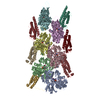
| |||||||||||||||||||||||||||||||||||||||||||||||||||||||||||||||||||||||||||||||||||||||||||||||||||||||||||||||||||||||||||||||||||||||||||||
|---|---|---|---|---|---|---|---|---|---|---|---|---|---|---|---|---|---|---|---|---|---|---|---|---|---|---|---|---|---|---|---|---|---|---|---|---|---|---|---|---|---|---|---|---|---|---|---|---|---|---|---|---|---|---|---|---|---|---|---|---|---|---|---|---|---|---|---|---|---|---|---|---|---|---|---|---|---|---|---|---|---|---|---|---|---|---|---|---|---|---|---|---|---|---|---|---|---|---|---|---|---|---|---|---|---|---|---|---|---|---|---|---|---|---|---|---|---|---|---|---|---|---|---|---|---|---|---|---|---|---|---|---|---|---|---|---|---|---|---|---|---|---|
| 1 |
| |||||||||||||||||||||||||||||||||||||||||||||||||||||||||||||||||||||||||||||||||||||||||||||||||||||||||||||||||||||||||||||||||||||||||||||
| Noncrystallographic symmetry (NCS) | NCS domain:
NCS domain segments:
|