- EMDB-22650: Cryo-EM structure of STRIPAK complex -
+
Open data
ID or keywords:
Loading...
-
Basic information
Entry
Database: EMDB / ID: EMD-22650
Title
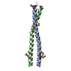
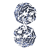
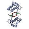
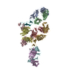
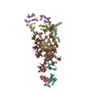
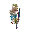
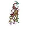
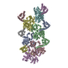
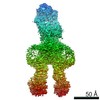
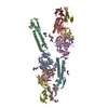
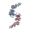
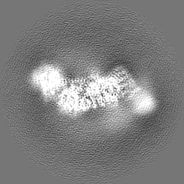
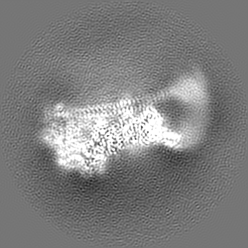
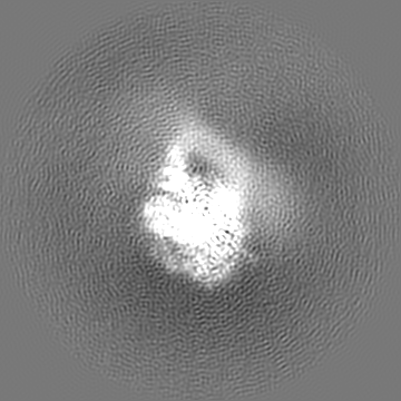
Cryo-EM structure of STRIPAK complex
Map data
SK7 primary map
Sample
Complex: SK7 complex
Protein or peptide: Serine/threonine-protein phosphatase 2A 65 kDa regulatory subunit A alpha isoform
Protein or peptide: Striatin-3
Protein or peptide: Serine/threonine-protein phosphatase 2A catalytic subunit alpha isoform
Protein or peptide: MOB-like protein phocein
Protein or peptide: Striatin-interacting protein 1
Ligand: MANGANESE (II) ION
Ligand: ZINC ION
Ligand: INOSITOL HEXAKISPHOSPHATEPhytic acid
Keywords
phosphorylation / complex / PP2A / SIGNALING PROTEIN
Function / homology
Function and homology information
FAR/SIN/STRIPAK complex / armadillo repeat domain binding / meiotic spindle elongation / Integration of energy metabolism / PP2A-mediated dephosphorylation of key metabolic factors / regulation of microtubule binding / MASTL Facilitates Mitotic Progression / mitotic sister chromatid separation / regulation of meiotic cell cycle process involved in oocyte maturation / protein phosphatase type 2A complex ...FAR/SIN/STRIPAK complex / armadillo repeat domain binding / meiotic spindle elongation / Integration of energy metabolism / PP2A-mediated dephosphorylation of key metabolic factors / regulation of microtubule binding / MASTL Facilitates Mitotic Progression / mitotic sister chromatid separation / regulation of meiotic cell cycle process involved in oocyte maturation / protein phosphatase type 2A complex / meiotic sister chromatid cohesion, centromeric / negative regulation of intracellular estrogen receptor signaling pathway / peptidyl-serine dephosphorylation / peptidyl-threonine dephosphorylation / : / positive regulation of microtubule binding / negative regulation of tyrosine phosphorylation of STAT protein / Regulation of glycolysis by fructose 2,6-bisphosphate metabolism / Inhibition of replication initiation of damaged DNA by RB1/E2F1 / female meiotic nuclear division / protein antigen binding / protein phosphatase regulator activity / ceramide metabolic process / GABA receptor binding / negative regulation of epithelial to mesenchymal transition / APC truncation mutants have impaired AXIN binding / AXIN missense mutants destabilize the destruction complex / Truncations of AMER1 destabilize the destruction complex / Initiation of Nuclear Envelope (NE) Reformation / ERKs are inactivated / positive regulation of extrinsic apoptotic signaling pathway in absence of ligand / Beta-catenin phosphorylation cascade / Signaling by GSK3beta mutants / CTNNB1 S33 mutants aren't phosphorylated / CTNNB1 S37 mutants aren't phosphorylated / CTNNB1 S45 mutants aren't phosphorylated / CTNNB1 T41 mutants aren't phosphorylated / regulation of cell morphogenesis / regulation of Wnt signaling pathway / Disassembly of the destruction complex and recruitment of AXIN to the membrane / regulation of growth / cortical actin cytoskeleton organization / negative regulation of glycolytic process through fructose-6-phosphate / Golgi cisterna membrane / positive regulation of NLRP3 inflammasome complex assembly / myosin phosphatase activity / protein serine/threonine phosphatase activity / CTLA4 inhibitory signaling / Platelet sensitization by LDL / negative regulation of MAPK cascade / protein-serine/threonine phosphatase / regulation of cell differentiation / T cell homeostasis / ERK/MAPK targets / regulation of G1/S transition of mitotic cell cycle / phosphoprotein phosphatase activity / regulation of DNA replication / mesoderm development / chromosome, centromeric region / DARPP-32 events / negative regulation of phosphatidylinositol 3-kinase/protein kinase B signal transduction / lateral plasma membrane / Nonsense Mediated Decay (NMD) enhanced by the Exon Junction Complex (EJC) / Amplification of signal from unattached kinetochores via a MAD2 inhibitory signal / regulation of cell adhesion / Cyclin A/B1/B2 associated events during G2/M transition / Mitotic Prometaphase / EML4 and NUDC in mitotic spindle formation / Loss of Nlp from mitotic centrosomes / Loss of proteins required for interphase microtubule organization from the centrosome / Recruitment of mitotic centrosome proteins and complexes / cytoskeleton organization / Resolution of Sister Chromatid Cohesion / Recruitment of NuMA to mitotic centrosomes / Anchoring of the basal body to the plasma membrane / AURKA Activation by TPX2 / protein dephosphorylation / RNA splicing / meiotic cell cycle / protein phosphatase 2A binding / response to organic substance / protein tyrosine phosphatase activity / chromosome segregation / RHO GTPases Activate Formins / response to lead ion / regulation of protein phosphorylation / Spry regulation of FGF signaling / RAF activation / PKR-mediated signaling / Degradation of beta-catenin by the destruction complex / tau protein binding / positive regulation of protein serine/threonine kinase activity / negative regulation of cell growth / small GTPase binding / kinase binding / spindle pole / Negative regulation of MAPK pathway / Separation of Sister Chromatids / Cyclin D associated events in G1 / microtubule cytoskeleton Similarity search - Function
Far11/STRP, N-terminal / Far11/STRP, C-terminal / Far11/STRP / N1221-like protein / Domain of unknown function (DUF3402) / N1221-like protein / Domain of unknown function (DUF3402) / Striatin, N-terminal / Striatin family / MOB kinase activator family ...Far11/STRP, N-terminal / Far11/STRP, C-terminal / Far11/STRP / N1221-like protein / Domain of unknown function (DUF3402) / N1221-like protein / Domain of unknown function (DUF3402) / Striatin, N-terminal / Striatin family / MOB kinase activator family / MOB kinase activator superfamily / Mob1/phocein family / Mob1/phocein family / : / HEAT repeat / HEAT repeat / Serine/threonine specific protein phosphatases signature. / Protein phosphatase 2A homologues, catalytic domain. / Serine/threonine-specific protein phosphatase/bis(5-nucleosyl)-tetraphosphatase / HEAT repeat profile. / HEAT, type 2 / HEAT repeats / Calcineurin-like phosphoesterase domain, ApaH type / Calcineurin-like phosphoesterase / Metallo-dependent phosphatase-like / Armadillo-like helical / Armadillo-type fold / G-protein beta WD-40 repeat / WD40 repeat, conserved site / Trp-Asp (WD) repeats signature. / WD domain, G-beta repeat / WD40 repeats / WD40 repeat / Trp-Asp (WD) repeats profile. / Trp-Asp (WD) repeats circular profile. / WD40-repeat-containing domain superfamily / WD40/YVTN repeat-like-containing domain superfamily Similarity search - Domain/homology
National Institutes of Health/National Institute of General Medical Sciences (NIH/NIGMS)
GM132275
United States
National Institutes of Health/National Institute of General Medical Sciences (NIH/NIGMS)
CA220283
United States
Welch Foundation
I-1932
United States
Welch Foundation
I-1944
United States
Welch Foundation
I-1702
United States
Citation
Journal: Nat Struct Mol Biol / Year: 2021 Title: Cryo-EM structure of the Hippo signaling integrator human STRIPAK. Authors: Byung-Cheon Jeong / Sung Jun Bae / Lisheng Ni / Xuewu Zhang / Xiao-Chen Bai / Xuelian Luo / Abstract: The striatin-interacting phosphatase and kinase (STRIPAK) complex is a large, multisubunit protein phosphatase 2A (PP2A) assembly that integrates diverse cellular signals in the Hippo pathway to ...The striatin-interacting phosphatase and kinase (STRIPAK) complex is a large, multisubunit protein phosphatase 2A (PP2A) assembly that integrates diverse cellular signals in the Hippo pathway to regulate cell proliferation and survival. The architecture and assembly mechanism of this critical complex are poorly understood. Using cryo-EM, we determine the structure of the human STRIPAK core comprising PP2AA, PP2AC, STRN3, STRIP1, and MOB4 at 3.2-Å resolution. Unlike the canonical trimeric PP2A holoenzyme, STRIPAK contains four copies of STRN3 and one copy of each the PP2AA-C heterodimer, STRIP1, and MOB4. The STRN3 coiled-coil domains form an elongated homotetrameric scaffold that links the complex together. An inositol hexakisphosphate (IP) is identified as a structural cofactor of STRIP1. Mutations of key residues at subunit interfaces disrupt the integrity of STRIPAK, causing aberrant Hippo pathway activation. Thus, STRIPAK is established as a noncanonical PP2A complex with four copies of regulatory STRN3 for enhanced signal integration.
History
Deposition
Sep 10, 2020
-
Header (metadata) release
Mar 10, 2021
-
Map release
Mar 10, 2021
-
Update
Mar 6, 2024
-
Current status
Mar 6, 2024
Processing site: RCSB / Status: Released
-
Structure visualization
Movie
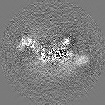
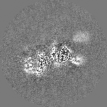
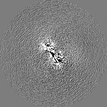
Surface view with section colored by density value
Model: Quantifoil R1.2/1.3 / Material: GOLD / Mesh: 300 / Support film - Material: CARBON / Support film - topology: HOLEY / Pretreatment - Type: GLOW DISCHARGE / Pretreatment - Time: 80 sec. / Pretreatment - Atmosphere: AIR / Details: The grid was coated with gold prior to use
Vitrification
Cryogen name: ETHANE / Chamber humidity: 100 % / Chamber temperature: 277 K / Instrument: FEI VITROBOT MARK IV
-
Electron microscopy
Microscope
FEI TITAN KRIOS
Electron beam
Acceleration voltage: 300 kV / Electron source: FIELD EMISSION GUN
In the structure databanks used in Yorodumi, some data are registered as the other names, "COVID-19 virus" and "2019-nCoV". Here are the details of the virus and the list of structure data.
Jan 31, 2019. EMDB accession codes are about to change! (news from PDBe EMDB page)
EMDB accession codes are about to change! (news from PDBe EMDB page)
The allocation of 4 digits for EMDB accession codes will soon come to an end. Whilst these codes will remain in use, new EMDB accession codes will include an additional digit and will expand incrementally as the available range of codes is exhausted. The current 4-digit format prefixed with “EMD-” (i.e. EMD-XXXX) will advance to a 5-digit format (i.e. EMD-XXXXX), and so on. It is currently estimated that the 4-digit codes will be depleted around Spring 2019, at which point the 5-digit format will come into force.
The EM Navigator/Yorodumi systems omit the EMD- prefix.
Related info.:Q: What is EMD? / ID/Accession-code notation in Yorodumi/EM Navigator
Yorodumi is a browser for structure data from EMDB, PDB, SASBDB, etc.
This page is also the successor to EM Navigator detail page, and also detail information page/front-end page for Omokage search.
The word "yorodu" (or yorozu) is an old Japanese word meaning "ten thousand". "mi" (miru) is to see.
Related info.:EMDB / PDB / SASBDB / Comparison of 3 databanks / Yorodumi Search / Aug 31, 2016. New EM Navigator & Yorodumi / Yorodumi Papers / Jmol/JSmol / Function and homology information / Changes in new EM Navigator and Yorodumi
 Movie
Movie Controller


 Open data
 Basic information
 Map data
 Sample
 Keywords
 phosphorylation /
  Function and homology information
 Authors
 United States, 5 items
  Citation
 Structure visualization
 Downloads & links
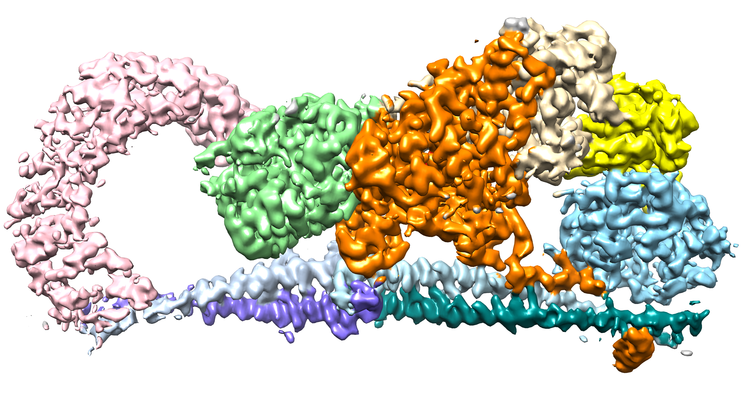 emd_22650.png
 http://ftp.pdbj.org/pub/emdb/structures/EMD-22650
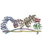


 Z
Z Y
Y X
X

 Sample components

 Processing
 Electron microscopy