 Movie
Movie Controller

[English] 日本語
 Yorodumi
- EMDB-2105: Single particle electron microscopy of PilQ dodecameric complexes... -
+ Open data

- Basic information
| Entry | Database: EMDB / ID: EMD-2105 | |||||||||
|---|---|---|---|---|---|---|---|---|---|---|
| Title | Single particle electron microscopy of PilQ dodecameric complexes from Neisseria meningitidis. | |||||||||
 Map data Map data | Reconstruction of outer membrane protein complex PilQ. | |||||||||
 Sample Sample |
| |||||||||
 Keywords Keywords |  Outer membrane protein / PilQ / pilus biogenesis Outer membrane protein / PilQ / pilus biogenesis | |||||||||
| Function / homology |  Function and homology information Function and homology informationtransport / establishment of competence for transformation / protein secretion / cell outer membrane / protein transport / membraneSimilarity search - Function | |||||||||
| Biological species |  Neisseria meningitidis MC58 (bacteria) Neisseria meningitidis MC58 (bacteria) | |||||||||
| Method | single particle reconstruction / cryo EM / negative staining / Resolution: 26.0 Å | |||||||||
 Authors Authors | Berry JL / Phelan MM / Collins RF / Adomavicius T / Tonjum T / Frye S / Bird L / Owens R / Ford RC / Lian LY / Derrick JP | |||||||||
 Citation Citation | Journal: PLoS Pathog / Year: 2012 Title: Structure and assembly of a trans-periplasmic channel for type IV pili in Neisseria meningitidis. Authors: Jamie-Lee Berry / Marie M Phelan / Richard F Collins / Tomas Adomavicius / Tone Tønjum / Stefan A Frye / Louise Bird / Ray Owens / Robert C Ford / Lu-Yun Lian / Jeremy P Derrick /  Abstract: Type IV pili are polymeric fibers which protrude from the cell surface and play a critical role in adhesion and invasion by pathogenic bacteria. The secretion of pili across the periplasm and outer ...Type IV pili are polymeric fibers which protrude from the cell surface and play a critical role in adhesion and invasion by pathogenic bacteria. The secretion of pili across the periplasm and outer membrane is mediated by a specialized secretin protein, PilQ, but the way in which this large channel is formed is unknown. Using NMR, we derived the structures of the periplasmic domains from N. meningitidis PilQ: the N-terminus is shown to consist of two β-domains, which are unique to the type IV pilus-dependent secretins. The structure of the second β-domain revealed an eight-stranded β-sandwich structure which is a novel variant of the HSP20-like fold. The central part of PilQ consists of two α/β fold domains: the structure of the first of these is similar to domains from other secretins, but with an additional α-helix which links it to the second α/β domain. We also determined the structure of the entire PilQ dodecamer by cryoelectron microscopy: it forms a cage-like structure, enclosing a cavity which is approximately 55 Å in internal diameter at its largest extent. Specific regions were identified in the density map which corresponded to the individual PilQ domains: this allowed us to dock them into the cryoelectron microscopy density map, and hence reconstruct the entire PilQ assembly which spans the periplasm. We also show that the C-terminal domain from the lipoprotein PilP, which is essential for pilus assembly, binds specifically to the first α/β domain in PilQ and use NMR chemical shift mapping to generate a model for the PilP:PilQ complex. We conclude that passage of the pilus fiber requires disassembly of both the membrane-spanning and the β-domain regions in PilQ, and that PilP plays an important role in stabilising the PilQ assembly during secretion, through its anchorage in the inner membrane. | |||||||||
| History |
|
- Structure visualization
| Movie |
Movie viewer |
|---|---|
| Structure viewer | EM map: SurfViewMolmilJmol/JSmol |
| Supplemental images |

- Downloads & links
-EMDB archive
| Map data | emd_2105.map.gz | 98.9 KB | EMDB map data format | |
|---|---|---|---|---|
| Header (meta data) | emd-2105-v30.xmlemd-2105.xml | 11.8 KB 11.8 KB | Display Display | EMDB header |
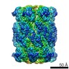
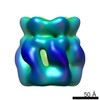
| Images | 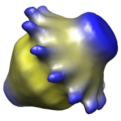 image2105.jpg image2105.jpg | 29.5 KB | ||
| Archive directory |  http://ftp.pdbj.org/pub/emdb/structures/EMD-2105ftp://ftp.pdbj.org/pub/emdb/structures/EMD-2105 http://ftp.pdbj.org/pub/emdb/structures/EMD-2105ftp://ftp.pdbj.org/pub/emdb/structures/EMD-2105 | HTTPS FTP |
-Related structure data
| Related structure data |  4av2MC  4aqzC  4ar0C M: atomic model generated by this map C: citing same article ( |
|---|---|
| Similar structure data |
-Links
| EMDB pages | EMDB (EBI/PDBe) / EMDataResource |
|---|---|
| Related items in Molecule of the Month |


-Map
| File | Download / File: emd_2105.map.gz / Format: CCP4 / Size: 1001 KB / Type: IMAGE STORED AS FLOATING POINT NUMBER (4 BYTES) | ||||||||||||||||||||||||||||||||||||||||||||||||||||||||||||||||||||
|---|---|---|---|---|---|---|---|---|---|---|---|---|---|---|---|---|---|---|---|---|---|---|---|---|---|---|---|---|---|---|---|---|---|---|---|---|---|---|---|---|---|---|---|---|---|---|---|---|---|---|---|---|---|---|---|---|---|---|---|---|---|---|---|---|---|---|---|---|---|
| Annotation | Reconstruction of outer membrane protein complex PilQ. | ||||||||||||||||||||||||||||||||||||||||||||||||||||||||||||||||||||
| Voxel size | X=Y=Z: 4.53 Å | ||||||||||||||||||||||||||||||||||||||||||||||||||||||||||||||||||||
| Density |
| ||||||||||||||||||||||||||||||||||||||||||||||||||||||||||||||||||||
| Symmetry | Space group: 1 | ||||||||||||||||||||||||||||||||||||||||||||||||||||||||||||||||||||
| Details | EMDB XML:
CCP4 map header:
| ||||||||||||||||||||||||||||||||||||||||||||||||||||||||||||||||||||
-Supplemental data
- Sample components
-Entire : Outer membrane protein PilQ from Neisseria meningitidis
| Entire | Name: Outer membrane protein PilQ from Neisseria meningitidis |
|---|---|
| Components |
|
-Supramolecule #1000: Outer membrane protein PilQ from Neisseria meningitidis
| Supramolecule | Name: Outer membrane protein PilQ from Neisseria meningitidis type: sample / ID: 1000 / Details: Isolated from Neisserial membranes. / Oligomeric state: Dodecamer / Number unique components: 1 |
|---|---|
| Molecular weight | Experimental: 960 KDa / Theoretical: 960 KDa / Method: From sequence |
-Macromolecule #1: PilQ
| Macromolecule | Name: PilQ / type: protein_or_peptide / ID: 1 / Number of copies: 1 / Oligomeric state: Dodecamer / Recombinant expression: No |
|---|---|
| Source (natural) | Organism: Neisseria meningitidis MC58 (bacteria) / Location in cell: Outer membrane |
| Molecular weight | Experimental: 960 KDa / Theoretical: 960 KDa |
| Sequence | GO: transport, protein secretion, protein transport, establishment of competence for transformation, cell outer membrane, membrane InterPro: AMIN domain, GspD/PilQ family, NolW-like, Type IV pilus secretin PilQ, Secretin/TonB, short N-terminal domain, Type II/III secretion system, Type II secretion system protein GspD, conserved site |
-Experimental details
-Structure determination
| Method | negative staining, cryo EM |
|---|---|
 Processing Processing | single particle reconstruction |
| Aggregation state | particle |
-Sample preparation
| Concentration | 1.0 mg/mL |
|---|---|
| Buffer | pH: 7.5 Details: 10 mM Tris-HCl, pH 7.5, 150 mM NaCl, 5 mM EDTA, and 0.1% (w/v) Zwittergent 3-10 |
| Staining | Type: NEGATIVE Details: Unstained: Both sides of the grid were briefly blotted dry with Whatman No. 1 filter paper (2 x 1s blots) using a Vitrobot (FEI) device (90% relative humidity), and the grid was then ...Details: Unstained: Both sides of the grid were briefly blotted dry with Whatman No. 1 filter paper (2 x 1s blots) using a Vitrobot (FEI) device (90% relative humidity), and the grid was then immediately plunged into liquid ethane maintained at <100K. |
| Grid | Details: 400 mesh Quantifoil holey carbon grid with 1.2 micron diameter holes. |
| Vitrification | Cryogen name: ETHANE / Chamber humidity: 90 % / Chamber temperature: 100 K / Instrument: FEI VITROBOT MARK III Method: Both sides of the grid were briefly blotted dry with Whatman No. 1 filter paper in a humidity-controlled chamber using a Vitrobot (FEI) device (90% relative humidity), and the grid was then ...Method: Both sides of the grid were briefly blotted dry with Whatman No. 1 filter paper in a humidity-controlled chamber using a Vitrobot (FEI) device (90% relative humidity), and the grid was then plunged into liquid ethane maintained at <100K. |
- Electron microscopy
| Microscope | FEI TECNAI F20 |
|---|---|
| Electron beam | Acceleration voltage: 200 kV / Electron source: FIELD EMISSION GUN |
| Electron optics | Calibrated magnification: 33112 / Illumination mode: FLOOD BEAM / Imaging mode: BRIGHT FIELDBright-field microscopy / Cs: 2.0 mm / Nominal defocus max: 5.1 µm / Nominal defocus min: 1.2 µm / Nominal magnification: 33000 |
| Sample stage | Specimen holder: Liquid nitrogen cooled / Specimen holder model: GATAN LIQUID NITROGEN |
| Temperature | Min: 94 K / Max: 100 K / Average: 97 K |
| Alignment procedure | Legacy - Astigmatism: Via FFT |
| Date | Nov 27, 2007 |
| Image recording | Category: CCD / Film or detector model: GENERIC GATAN (4k x 4k) / Number real images: 55 / Average electron dose: 4 e/Å2 / Bits/pixel: 8 |
| Experimental equipment |  Model: Tecnai F20 / Image courtesy: FEI Company |
-Image processing
| CTF correction | Details: CTFFIT each micrograph |
|---|---|
| Final two d classification | Number classes: 101 |
| Final reconstruction | Applied symmetry - Point group: C12 (12 fold cyclic) / Algorithm: OTHER / Resolution.type: BY AUTHOR / Resolution: 26.0 Å / Resolution method: OTHER / Software - Name: EMAN Details: Two different starting models were employed with convergence between them. This map was generated from model 1. Number images used: 25303 |
| Details | 29000 |
