 Movie
Movie Controller

[English] 日本語
 Yorodumi
- PDB-1dgi: Cryo-EM structure of human poliovirus(serotype 1)complexed with t... -
+ Open data

- Basic information
| Entry | Database: PDB / ID: 1dgi | ||||||
|---|---|---|---|---|---|---|---|
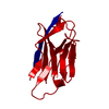
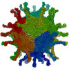
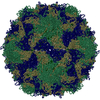
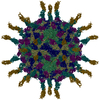
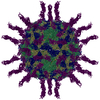
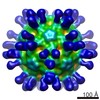
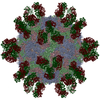
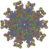
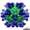
| Title | Cryo-EM structure of human poliovirus(serotype 1)complexed with three domain CD155 | ||||||
 Components Components |
| ||||||
 Keywords Keywords | Virus/Receptor / CD155 / PVR / HUMAN POLIOVIRUS / POLIOVIRUS-RECEPTOR COMPLEX / Icosahedral virus / Virus-Receptor COMPLEX | ||||||
| Function / homology |  Function and homology information Function and homology informationsusceptibility to T cell mediated cytotoxicity / susceptibility to natural killer cell mediated cytotoxicity / Nectin/Necl trans heterodimerization / positive regulation of natural killer cell mediated cytotoxicity directed against tumor cell target / symbiont-mediated suppression of host translation initiation / positive regulation of natural killer cell mediated cytotoxicity / heterophilic cell-cell adhesion via plasma membrane cell adhesion molecules / homophilic cell adhesion via plasma membrane adhesion molecules / symbiont-mediated suppression of host cytoplasmic pattern recognition receptor signaling pathway via inhibition of RIG-I activity / symbiont-mediated suppression of host cytoplasmic pattern recognition receptor signaling pathway via inhibition of MDA-5 activity ...susceptibility to T cell mediated cytotoxicity / susceptibility to natural killer cell mediated cytotoxicity / Nectin/Necl trans heterodimerization / positive regulation of natural killer cell mediated cytotoxicity directed against tumor cell target / symbiont-mediated suppression of host translation initiation / positive regulation of natural killer cell mediated cytotoxicity / heterophilic cell-cell adhesion via plasma membrane cell adhesion molecules / homophilic cell adhesion via plasma membrane adhesion molecules / symbiont-mediated suppression of host cytoplasmic pattern recognition receptor signaling pathway via inhibition of RIG-I activity / symbiont-mediated suppression of host cytoplasmic pattern recognition receptor signaling pathway via inhibition of MDA-5 activity / cell adhesion molecule binding / symbiont-mediated suppression of host cytoplasmic pattern recognition receptor signaling pathway via inhibition of MAVS activity / picornain 2A / symbiont-mediated suppression of host mRNA export from nucleus / ribonucleoside triphosphate phosphatase activity / symbiont genome entry into host cell via pore formation in plasma membrane / picornain 3C / T=pseudo3 icosahedral viral capsid / adherens junction / host cell cytoplasmic vesicle membrane / endocytosis involved in viral entry into host cell / Immunoregulatory interactions between a Lymphoid and a non-Lymphoid cell / virus receptor activity / nucleoside-triphosphate phosphatase / protein complex oligomerization / monoatomic ion channel activity / signaling receptor activity / RNA helicase activity / induction by virus of host autophagy / cysteine-type endopeptidase activity / RNA-directed RNA polymerase / focal adhesion / viral RNA genome replication / virus-mediated perturbation of host defense response / RNA-dependent RNA polymerase activity / DNA-templated transcription / host cell nucleus / virion attachment to host cell / structural molecule activity / cell surface / proteolysis / RNA binding / extracellular space / ATP binding / membrane / metal ion binding / plasma membrane / cytoplasm Similarity search - Function | ||||||
| Biological species |  Homo sapiens (human) Homo sapiens (human) Human poliovirus 1 Human poliovirus 1 | ||||||
| Method | ELECTRON MICROSCOPY / single particle reconstruction / cryo EM / Resolution: 22 Å | ||||||
 Authors Authors | He, Y. / Bowman, V.D. / Mueller, S. / Bator, C.M. / Bella, J. / Peng, X. / Baker, T.S. / Wimmer, E. / Kuhn, R.J. / Rossmann, M.G. | ||||||
 Citation Citation | Journal: Proc Natl Acad Sci U S A / Year: 2000 Title: Interaction of the poliovirus receptor with poliovirus. Authors: Y He / V D Bowman / S Mueller / C M Bator / J Bella / X Peng / T S Baker / E Wimmer / R J Kuhn / M G Rossmann /  Abstract: The structure of the extracellular, three-domain poliovirus receptor (CD155) complexed with poliovirus (serotype 1) has been determined to 22-A resolution by means of cryo-electron microscopy and ...The structure of the extracellular, three-domain poliovirus receptor (CD155) complexed with poliovirus (serotype 1) has been determined to 22-A resolution by means of cryo-electron microscopy and three-dimensional image-reconstruction techniques. Density corresponding to the receptor was isolated in a difference electron density map and fitted with known structures, homologous to those of the three individual CD155 Ig-like domains. The fit was confirmed by the location of carbohydrate moieties in the CD155 glycoprotein, the conserved properties of elbow angles in the structures of cell surface molecules with Ig-like folds, and the concordance with prior results of CD155 and poliovirus mutagenesis. CD155 binds in the poliovirus "canyon" and has a footprint similar to that of the intercellular adhesion molecule-1 receptor on human rhinoviruses. However, the orientation of the long, slender CD155 molecule relative to the poliovirus surface is quite different from the orientation of intercellular adhesion molecule-1 on rhinoviruses. In addition, the residues that provide specificity of recognition differ for the two receptors. The principal feature of receptor binding common to these two picornaviruses is the site in the canyon at which binding occurs. This site may be a trigger for initiation of the subsequent uncoating step required for viral infection. #1: Journal: Proc.Natl.Acad.Sci.USA / Year: 1998Title: The Structure of the Two Amino-Terminal Domain of Human Icam-1 Suggests How It Functions as a Rhinovirus Receptor and as an Lfa-1 Integrin Ligand Authors: Bella, J. / Kolatkar, P. / Marlor, C.W. / Greve, J. / Rossmann, M.G. #2: Journal: Science / Year: 1985Title: Three-Dimensional Structure of Poliovirus at 2.9A Resolution Authors: Hogle, J. / Chow, M. / Filman, D.J. | ||||||
| History |
|
- Structure visualization
| Movie |
Movie viewer |
|---|---|
| Structure viewer | Molecule: MolmilJmol/JSmol |
- Downloads & links
-Download
| PDBx/mmCIF format | 1dgi.cif.gz | 61 KB | Display | PDBx/mmCIF format |
|---|---|---|---|---|
| PDB format | pdb1dgi.ent.gz | 30.4 KB | Display | PDB format |
| PDBx/mmJSON format | 1dgi.json.gz | Tree view | PDBx/mmJSON format | |
| Others |  Other downloads Other downloads |
-Validation report
| Summary document | 1dgi_validation.pdf.gz | 320 KB | Display | wwPDB validaton report |
|---|---|---|---|---|
| Full document | 1dgi_full_validation.pdf.gz | 319.5 KB | Display | |
| Data in XML | 1dgi_validation.xml.gz | 1011 B | Display | |
| Data in CIF | 1dgi_validation.cif.gz | 11.4 KB | Display | |
| Arichive directory | https://data.pdbj.org/pub/pdb/validation_reports/dg/1dgiftp://data.pdbj.org/pub/pdb/validation_reports/dg/1dgi | HTTPS FTP |
-Related structure data
| Related structure data | |
|---|---|
| Similar structure data |


-Links
 PDBj
PDBj


- Assembly
| Deposited unit | 
|
|---|---|
| 1 | x 60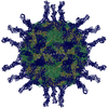
|
| 2 |
|
| 3 | x 5
|
| 4 | x 6
|
| 5 | 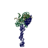
|
| Symmetry | Point symmetry: (Hermann–Mauguin notation: 532 / Schoenflies symbol: I (icosahedral)) |
-Components
| #1: Protein | Mass: 32928.160 Da / Num. of mol.: 1 / Fragment: THREE EXTRACELLULAR DOMAINS OF CD155 Source method: isolated from a genetically manipulated source Source: (gene. exp.) Homo sapiens (human) / Cell (production host): KIDNEY CELLS / Production host: Homo sapiens (human) / References: UniProt: P15151 |
|---|---|
| #2: Protein | Mass: 31874.705 Da / Num. of mol.: 1 Source method: isolated from a genetically manipulated source Source: (gene. exp.) Human poliovirus 1 / Genus: Enterovirus / Species: Poliovirus / Cell (production host): HELA CELLS / Production host: Homo sapiens (human) / References: UniProt: P03300 |
| #3: Protein | Mass: 29664.332 Da / Num. of mol.: 1 Source method: isolated from a genetically manipulated source Source: (gene. exp.) Human poliovirus 1 / Genus: Enterovirus / Species: Poliovirus / Cell (production host): HELA CELLS / Production host: Homo sapiens (human) / References: UniProt: P03300 |
| #4: Protein | Mass: 26235.115 Da / Num. of mol.: 1 Source method: isolated from a genetically manipulated source Source: (gene. exp.) Human poliovirus 1 / Genus: Enterovirus / Species: Poliovirus / Cell (production host): HELA CELLS / Production host: Homo sapiens (human) / References: UniProt: P03300 |
| #5: Protein | Mass: 6983.754 Da / Num. of mol.: 1 / Fragment: POLIOVIRUS FRAGMENTS VP1,VP2,VP3,VP4 Source method: isolated from a genetically manipulated source Source: (gene. exp.) Human poliovirus 1 / Genus: Enterovirus / Species: Poliovirus / Cell (production host): HELA CELLS / Production host: Homo sapiens (human) / References: UniProt: P03300*PLUS |
-Experimental details
-Experiment
| Experiment | Method: ELECTRON MICROSCOPY |
|---|---|
| EM experiment | Aggregation state: PARTICLE / 3D reconstruction method: single particle reconstruction |
- Sample preparation
| Component | Name: human poliovirus(serotype 1)complexed with three domain CD155 Type: COMPLEX |
|---|---|
| Buffer solution | pH: 7.5 |
| Specimen | Embedding applied: NO / Shadowing applied: NO / Staining applied: NO / Vitrification applied: YES |
| Vitrification | Details: POLIOVIRUS WAS INCUBATED WITH CD155-AP FOR 1 HOURS AT 4 DEGREES CELSIUS (277 KELVIN) USING A EIGHT-FOLD EXCESS OF CD155-AP FOR EACH OF THE SIXTY POSSIBLE BINDING SITES PER VIRION. AFTER ...Details: POLIOVIRUS WAS INCUBATED WITH CD155-AP FOR 1 HOURS AT 4 DEGREES CELSIUS (277 KELVIN) USING A EIGHT-FOLD EXCESS OF CD155-AP FOR EACH OF THE SIXTY POSSIBLE BINDING SITES PER VIRION. AFTER INCUBATION, SAMPLES WERE PREPARED AS THIN LAYERS OF VITREOUS ICE AND MAINTAINED AT NEAR LIQUID NITROGEN TEMPERATURE IN THE ELECTRON MICROSCOPE WITH A GATAN 626 CRYOTRANSFER HOLDER. |
| Crystal grow | pH: 7.5 Details: WARNING: THIS IS AN ELECTRON MICROSCOPY MODEL DEPOSITION. CRYO-EM INFORMATION HAS BEEN INCLUDED IN THE FORM OF REMARK 250 RECORDS AT THE TOP OF THE PDB COORDINATE FILE., pH 7.5, ELECTRON ...Details: WARNING: THIS IS AN ELECTRON MICROSCOPY MODEL DEPOSITION. CRYO-EM INFORMATION HAS BEEN INCLUDED IN THE FORM OF REMARK 250 RECORDS AT THE TOP OF THE PDB COORDINATE FILE., pH 7.5, ELECTRON MICROSCOPY RECONSTRUCTION |
| Crystal grow | *PLUS Method: unknown / Details: electron microscopy |
- Electron microscopy imaging
| Microscopy | Model: FEI/PHILIPS CM200FEG / Date: Jun 1, 1999 |
|---|---|
| Electron gun | Electron source:  FIELD EMISSION GUN / Accelerating voltage: 80 kV / Illumination mode: FLOOD BEAM FIELD EMISSION GUN / Accelerating voltage: 80 kV / Illumination mode: FLOOD BEAM |
| Electron lens | Mode: BRIGHT FIELD / Nominal magnification: 38000 X / Nominal defocus max: 2100 nm / Nominal defocus min: 1300 nm |
| Specimen holder | Temperature: 120 K |
| Image recording | Electron dose: 20 e/Å2 / Film or detector model: KODAK SO-163 FILM |
- Processing
| EM software | Name: PURDUE PROGRAMS / Category: 3D reconstruction | ||||||||||||
|---|---|---|---|---|---|---|---|---|---|---|---|---|---|
| Symmetry | Point symmetry: I (icosahedral) | ||||||||||||
| 3D reconstruction | Method: COMMON-LINES AND POLAR-FOURIER-TRANSFORM FULLER ET AL. 1996, J.STRUC.BIOL.c 116, 48-55; BAKER AND CHENG, 1996, J.STRUC.BIOL. 116, 120-130 Resolution: 22 Å / Resolution method: OTHER / Num. of particles: 1156 / Actual pixel size: 3.68 Å Magnification calibration: THE PIXEL SIZE OF THE CRYO-EM MAP WAS CALIBRATED AGAINST A LOW RESOLUTION DENSITY MAP CALCULATED FROM THE CRYSTAL STRUCTURE OF POLIOVIRUS. DENSITIES WERE COMPARED BY CROSS- ...Magnification calibration: THE PIXEL SIZE OF THE CRYO-EM MAP WAS CALIBRATED AGAINST A LOW RESOLUTION DENSITY MAP CALCULATED FROM THE CRYSTAL STRUCTURE OF POLIOVIRUS. DENSITIES WERE COMPARED BY CROSS- CORRELATION WITHIN A SPHERICAL SHELL OF INTERNAL RADIUS 110 ANGSTROMS AND EXTERNAL RADIUS OF 145 ANGSTROMS. Details: THE RESOLUTION OF THE FINAL RECONSTRUCTED DENSITY WAS DETERMINED TO BE AT LEAST 22 ANGSTROMS, AS MEASURED BY RANDOMLY SPLITTING THE PARTICLES INTO TWO SETS AND COMPARING STRUCTURE FACTORS ...Details: THE RESOLUTION OF THE FINAL RECONSTRUCTED DENSITY WAS DETERMINED TO BE AT LEAST 22 ANGSTROMS, AS MEASURED BY RANDOMLY SPLITTING THE PARTICLES INTO TWO SETS AND COMPARING STRUCTURE FACTORS OBTAINED FROM SEPARATE RECONSTRUCTIONS (BAKER ET AL. 1991, BIOPHYS.J. 60, 1445-1456). THE EIGENVALUE SPECTRUM GAVE AN INDICATION OF THE RANDOMNESS OF THE DATA THAT WAS INCLUDED IN THE RECONSTRUCTION. THE COMPLETENESS OF THE DATA WAS VERIFIED IN THAT ALL EIGENVALUES EXCEEDED 1.0. Symmetry type: POINT | ||||||||||||
| Atomic model building | Protocol: OTHER / Space: REAL / Target criteria: VISUAL AGREEMENT Details: METHOD--MANUAL REFINEMENT PROTOCOL--MANUAL ADJUSTMENT DETAILS--THE CRYSTAL STRUCTURE OF POLIOVIRUS WAS PLACED INTO THE CALIBRATED CRYO-EM DENSITY MAP BY ALIGNING THE ICOSAHEDRAL SYMMETRY ...Details: METHOD--MANUAL REFINEMENT PROTOCOL--MANUAL ADJUSTMENT DETAILS--THE CRYSTAL STRUCTURE OF POLIOVIRUS WAS PLACED INTO THE CALIBRATED CRYO-EM DENSITY MAP BY ALIGNING THE ICOSAHEDRAL SYMMETRY AXES. APPROPRIATELY GLYCOSYLATED MODELS OF CD155 WITH VARIOUS INTERDOMAIN ANGLES WERE MANUALLY FITTED INTO THE DIFFERENCE DENSITY CALCULAT BY SUBSTRACTING POLIOVIRUS NATIVE RECONSTRUCTION FROM THE COMPLEX RECONSTRUCTION. THE COORDINATES ARE IN THE P, Q, R FRAME IN ANGSTROM UNITS AND CORRESPOND TO ICOSAHEDRAL SYMMETRY AXES. THE ORIGIN IS CHOSEN AT THE CENTER OF THE VIRUS WITH P, Q AND R ALONG MUTUALLY PERPENDICULAR TWO-FOLD AXES OF THE ICOSAHEDRON. THEY SHOULD REMAIN IN THAT FRAME FOR THE EASE OF THE USER IN CREATING THE BIOLOGICALLY SIGNIFICANT VIRAL COMPLEX PARTICLE USING THE 60 ICOSAHEDRAL SYMMETRY OPERATORS. CD155 MODEL BUILDING (D1-D2-D3)-- ATOMIC MODEL OF D1 WAS BUILT BASED ON MYELIN PROTEIN ZERO(1NEU). ATOMIC MODEL OF D2 WAS BUILT FROM A FAB FRAGMENT(1CIC).ATOMIC MODEL OF D3 WAS BUILT FROM AN INSECT IMMUNE PROTEIN(1BIH).THE ELBOW ANGLE BETWEEN D1 AND D2 WAS DETERMINED BY LEAST-SQUARE-FITTING THESE TWO DOMAINS THE CRYSTAL STRUCTURE OF CD2(1HNF). | ||||||||||||
| Refinement | Highest resolution: 22 Å | ||||||||||||
| Refinement step | Cycle: LAST / Highest resolution: 22 Å
|
