Movie
Movie Controller

[English] 日本語
 Yorodumi
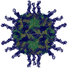
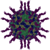
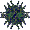
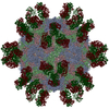
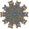
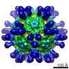
- PDB-3j9f: Poliovirus complexed with soluble, deglycosylated poliovirus rece... -
+ Open data

- Basic information
| Entry | Database: PDB / ID: 3j9f | |||||||||
|---|---|---|---|---|---|---|---|---|---|---|
| Title | Poliovirus complexed with soluble, deglycosylated poliovirus receptor (Pvr) at 4 degrees C | |||||||||
 Components Components |
| |||||||||
 Keywords Keywords | VIRUS/CELL ADHESION / deglycosylated receptor /  picornavirus / PVR / CD155 / enterovirus / cell entry / VIRUS-CELL ADHESION complex picornavirus / PVR / CD155 / enterovirus / cell entry / VIRUS-CELL ADHESION complex | |||||||||
| Function / homology |  Function and homology information Function and homology informationsusceptibility to T cell mediated cytotoxicity / susceptibility to natural killer cell mediated cytotoxicity / Nectin/Necl trans heterodimerization / positive regulation of natural killer cell mediated cytotoxicity directed against tumor cell target / symbiont-mediated suppression of host translation initiation / heterophilic cell-cell adhesion via plasma membrane cell adhesion molecules / positive regulation of natural killer cell mediated cytotoxicity / homophilic cell adhesion via plasma membrane adhesion molecules / symbiont-mediated suppression of host cytoplasmic pattern recognition receptor signaling pathway via inhibition of MDA-5 activity / symbiont-mediated suppression of host cytoplasmic pattern recognition receptor signaling pathway via inhibition of RIG-I activity ...susceptibility to T cell mediated cytotoxicity / susceptibility to natural killer cell mediated cytotoxicity / Nectin/Necl trans heterodimerization / positive regulation of natural killer cell mediated cytotoxicity directed against tumor cell target / symbiont-mediated suppression of host translation initiation / heterophilic cell-cell adhesion via plasma membrane cell adhesion molecules / positive regulation of natural killer cell mediated cytotoxicity / homophilic cell adhesion via plasma membrane adhesion molecules / symbiont-mediated suppression of host cytoplasmic pattern recognition receptor signaling pathway via inhibition of MDA-5 activity / symbiont-mediated suppression of host cytoplasmic pattern recognition receptor signaling pathway via inhibition of RIG-I activity / picornain 2A / symbiont-mediated suppression of host cytoplasmic pattern recognition receptor signaling pathway via inhibition of MAVS activity / symbiont-mediated suppression of host mRNA export from nucleus / symbiont genome entry into host cell via pore formation in plasma membrane / cell adhesion molecule binding / ribonucleoside triphosphate phosphatase activity / picornain 3C / T=pseudo3 icosahedral viral capsid / host cell cytoplasmic vesicle membrane / adherens junction / endocytosis involved in viral entry into host cell / : / Immunoregulatory interactions between a Lymphoid and a non-Lymphoid cell / nucleoside-triphosphate phosphatase / protein complex oligomerization / virus receptor activity / signaling receptor activity / monoatomic ion channel activity / RNA helicase activity / induction by virus of host autophagy / RNA-directed RNA polymerase / symbiont-mediated suppression of host gene expression / viral RNA genome replication / cysteine-type endopeptidase activity / RNA-dependent RNA polymerase activity / focal adhesion / DNA-templated transcription / host cell nucleus / structural molecule activity / virion attachment to host cell / cell surface / proteolysis / extracellular space / RNA binding / ATP binding / membrane / metal ion binding / plasma membrane / cytoplasmSimilarity search - Function | |||||||||
| Biological species |   Human poliovirus 1 Mahoney Human poliovirus 1 Mahoney Homo sapiens (human) Homo sapiens (human) | |||||||||
| Method | ELECTRON MICROSCOPY / single particle reconstruction / cryo EM / Resolution: 9 Å | |||||||||
 Authors Authors | Strauss, M. / Filman, D.J. / Belnap, D.M. / Cheng, N. / Noel, R.T. / Hogle, J.M. | |||||||||
 Citation Citation | Journal: J Virol / Year: 2015 Title: Nectin-like interactions between poliovirus and its receptor trigger conformational changes associated with cell entry. Authors: Mike Strauss / David J Filman / David M Belnap / Naiqian Cheng / Roane T Noel / James M Hogle /  Abstract: Poliovirus infection is initiated by attachment to a receptor on the cell surface called Pvr or CD155. At physiological temperatures, the receptor catalyzes an irreversible expansion of the virus to ...Poliovirus infection is initiated by attachment to a receptor on the cell surface called Pvr or CD155. At physiological temperatures, the receptor catalyzes an irreversible expansion of the virus to form an expanded form of the capsid called the 135S particle. This expansion results in the externalization of the myristoylated capsid protein VP4 and the N-terminal extension of the capsid protein VP1, both of which become inserted into the cell membrane. Structures of the expanded forms of poliovirus and of several related viruses have recently been reported. However, until now, it has been unclear how receptor binding triggers viral expansion at physiological temperature. Here, we report poliovirus in complex with an enzymatically partially deglycosylated form of the 3-domain ectodomain of Pvr at a 4-Å resolution, as determined by cryo-electron microscopy. The interaction of the receptor with the virus in this structure is reminiscent of the interactions of Pvr with its natural ligands. At a low temperature, the receptor induces very few changes in the structure of the virus, with the largest changes occurring within the footprint of the receptor, and in a loop of the internal protein VP4. Changes in the vicinity of the receptor include the displacement of a natural lipid ligand (called "pocket factor"), demonstrating that the loss of this ligand, alone, is not sufficient to induce particle expansion. Finally, analogies with naturally occurring ligand binding in the nectin family suggest which specific structural rearrangements in the virus-receptor complex could help to trigger the irreversible expansion of the capsid. IMPORTANCE: The cell-surface receptor (Pvr) catalyzes a large structural change in the virus that exposes membrane-binding protein chains. We fitted known atomic models of the virus and Pvr into ...IMPORTANCE: The cell-surface receptor (Pvr) catalyzes a large structural change in the virus that exposes membrane-binding protein chains. We fitted known atomic models of the virus and Pvr into three-dimensional experimental maps of the receptor-virus complex. The molecular interactions we see between poliovirus and its receptor are reminiscent of the nectin family, by involving the burying of otherwise-exposed hydrophobic groups. Importantly, poliovirus expansion is regulated by the binding of a lipid molecule within the viral capsid. We show that receptor binding either causes this molecule to be expelled or requires it, but that its loss is not sufficient to trigger irreversible expansion. Based on our model, we propose testable hypotheses to explain how the viral shell becomes destabilized, leading to RNA uncoating. These findings give us a better understanding of how poliovirus has evolved to exploit a natural process of its host to penetrate the membrane barrier. | |||||||||
| History |
|
- Structure visualization
| Movie |
Movie viewer |
|---|---|
| Structure viewer | Molecule: MolmilJmol/JSmol |
- Downloads & links
-Download
| PDBx/mmCIF format | 3j9f.cif.gz | 216.7 KB | Display | PDBx/mmCIF format |
|---|---|---|---|---|
| PDB format | pdb3j9f.ent.gz | 170.2 KB | Display | PDB format |
| PDBx/mmJSON format | 3j9f.json.gz | Tree view | PDBx/mmJSON format | |
| Others |  Other downloads Other downloads |
-Validation report
| Arichive directory | https://data.pdbj.org/pub/pdb/validation_reports/j9/3j9fftp://data.pdbj.org/pub/pdb/validation_reports/j9/3j9f | HTTPS FTP |
|---|
-Related structure data
| Related structure data | 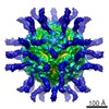 6243MC 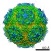 6147C  6148C 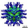 6242C 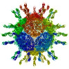 3j8fC M: map data used to model this data C: citing same article ( |
|---|---|
| Similar structure data |
-Links
 PDBj
PDBj


- Assembly
| Deposited unit | 
|
|---|---|
| 1 | x 60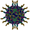
|
| 2 |
|
| 3 | x 5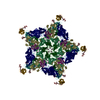
|
| 4 | x 6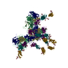
|
| 5 | 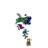
|
| Symmetry | Point symmetry: (Schoenflies symbol: I (icosahedral)) |
-Components
-Protein , 7 types, 7 molecules 1234789
| #1: Protein | Mass: 33488.613 Da / Num. of mol.: 1 / Fragment: UNP residues 580-881 / Source method: isolated from a natural source / Source: (natural) Human poliovirus 1 Mahoney / References: UniProt: P03300 |
|---|---|
| #2: Protein | Mass: 30075.783 Da / Num. of mol.: 1 / Fragment: UNP residues 70-341 / Source method: isolated from a natural source / Source: (natural) Human poliovirus 1 Mahoney / References: UniProt: P03300 |
| #3: Protein | Mass: 26547.482 Da / Num. of mol.: 1 / Fragment: UNP residues 342-579 / Source method: isolated from a natural source / Source: (natural) Human poliovirus 1 Mahoney / References: UniProt: P03300 |
| #4: Protein | Mass: 7603.405 Da / Num. of mol.: 1 / Fragment: UNP residues 2-69 / Source method: isolated from a natural source / Source: (natural) Human poliovirus 1 Mahoney / References: UniProt: P03300 |
| #5: Protein | CD155 / Nectin-like protein 5 / NECL-5 / PVR / CD155 Mass: 12869.564 Da / Num. of mol.: 1 / Fragment: SEE REMARK 999 / Source method: isolated from a natural source / Source: (natural) Homo sapiens (human) / References: UniProt: P15151 |
| #6: Protein | CD155 / Nectin-like protein 5 / NECL-5 / PVR / CD155 Mass: 10999.521 Da / Num. of mol.: 1 / Fragment: SEE REMARK 999 / Source method: isolated from a natural source / Source: (natural) Homo sapiens (human) / References: UniProt: P15151 |
| #7: Protein | CD155 / Nectin-like protein 5 / NECL-5 / PVR / CD155 Mass: 9952.081 Da / Num. of mol.: 1 / Fragment: SEE REMARK 999 / Source method: isolated from a natural source / Source: (natural) Homo sapiens (human) / References: UniProt: P15151 |
-Sugars , 5 types, 7 molecules 
| #8: Polysaccharide | / Mass: 586.542 Da / Num. of mol.: 2 Source method: isolated from a genetically manipulated source #9: Polysaccharide | alpha-D-mannopyranose-(1-3)-beta-D-mannopyranose-(1-4)-2-acetamido-2-deoxy-beta-D-glucopyranose-(1- ...alpha-D-mannopyranose-(1-3)-beta-D-mannopyranose-(1-4)-2-acetamido-2-deoxy-beta-D-glucopyranose-(1-4)-[alpha-L-fucopyranose-(1-6)]2-acetamido-2-deoxy-beta-D-glucopyranose | / Mass: 894.823 Da / Num. of mol.: 1Source method: isolated from a genetically manipulated source #10: Polysaccharide | / Mass: 424.401 Da / Num. of mol.: 2Source method: isolated from a genetically manipulated source #11: Polysaccharide | beta-D-mannopyranose-(1-4)-2-acetamido-2-deoxy-beta-D-glucopyranose-(1-4)-[alpha-L-fucopyranose-(1- ...beta-D-mannopyranose-(1-4)-2-acetamido-2-deoxy-beta-D-glucopyranose-(1-4)-[alpha-L-fucopyranose-(1-6)]2-acetamido-2-deoxy-beta-D-glucopyranose | / Mass: 732.682 Da / Num. of mol.: 1Source method: isolated from a genetically manipulated source #13: Sugar | ChemComp-NAG / | N-Acetylglucosamine Type: D-saccharide, beta linking / Mass: 221.208 Da / Num. of mol.: 1 Type: D-saccharide, beta linking / Mass: 221.208 Da / Num. of mol.: 1Source method: isolated from a genetically manipulated source Formula: C8H15NO6 |
|---|
-Non-polymers , 1 types, 1 molecules 
| #12: Chemical | ChemComp-PLM / Palmitic acid Mass: 256.424 Da / Num. of mol.: 1 / Source method: obtained synthetically / Formula: C16H32O2 Mass: 256.424 Da / Num. of mol.: 1 / Source method: obtained synthetically / Formula: C16H32O2 |
|---|
-Details
| Nonpolymer details | GLYCOSYLATION IN THIS ENTRY IS DERIVED FROM STARTING STRUCTURE PDB ENTRY 4FQP AND DOES NOT ...GLYCOSYLAT |
|---|---|
| Sequence details | THE RECEPTOR HAS BEEN MODELED AS THREE INDIVIDUAL DOMAINS WITH DUPLICATED, CLASHING LINKER SEGMENTS ...THE RECEPTOR HAS BEEN MODELED AS THREE INDIVIDUAL |
-Experimental details
-Experiment
| Experiment | Method: ELECTRON MICROSCOPY |
|---|---|
| EM experiment | Aggregation state: PARTICLE / 3D reconstruction method: single particle reconstruction |
- Sample preparation
| Component |
| ||||||||||||||||||||
|---|---|---|---|---|---|---|---|---|---|---|---|---|---|---|---|---|---|---|---|---|---|
| Details of virus | Empty: NO / Enveloped: NO / Host category: VERTEBRATES / Isolate: STRAIN / Type: VIRION | ||||||||||||||||||||
| Natural host | Organism: Homo sapiens | ||||||||||||||||||||
| Specimen | Embedding applied: NO / Shadowing applied: NO / Staining applied: NO / Vitrification applied: YES | ||||||||||||||||||||
| Vitrification | Instrument: HOMEMADE PLUNGER / Cryogen name: ETHANE |
- Electron microscopy imaging
| Microscopy | Model: FEI/PHILIPS CM200FEG / Date: Nov 1, 1999 |
|---|---|
| Electron gun | Electron source: FIELD EMISSION GUN / Accelerating voltage: 120 kV / Illumination mode: FLOOD BEAM |
| Electron lens | Mode: BRIGHT FIELDBright-field microscopy / Cs: 2 mm |
| Specimen holder | Specimen holder model: GATAN LIQUID NITROGEN |
| Image recording | Film or detector model: KODAK SO-163 FILM |
- Processing
| Symmetry | Point symmetry: I (icosahedral) | ||||||||||||
|---|---|---|---|---|---|---|---|---|---|---|---|---|---|
| 3D reconstruction | Resolution: 9 Å / Resolution method: FSC 0.5 CUT-OFF / Num. of particles: 3822 / Nominal pixel size: 1.77182 Å / Actual pixel size: 1.77182 Å / Details: (Single particle--Applied symmetry: I) / Symmetry type: POINT | ||||||||||||
| Atomic model building | Protocol: RIGID BODY FIT / Space: RECIPROCAL / Details: REFINEMENT PROTOCOL--rigid body | ||||||||||||
| Atomic model building | PDB-ID: 1HXS | ||||||||||||
| Refinement step | Cycle: LAST
|