 Movie
Movie Controller

[English] 日本語
 Yorodumi
- EMDB-1421: Electron cryomicroscopy reveals different F1+F2 protein States in... -
+ Open data

- Basic information
| Entry | Database: EMDB / ID: EMD-1421 | |||||||||
|---|---|---|---|---|---|---|---|---|---|---|
| Title | Electron cryomicroscopy reveals different F1+F2 protein States in intact parainfluenza virions. | |||||||||
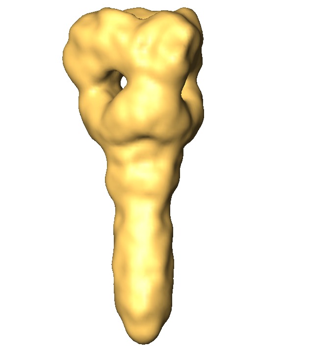
 Map data Map data | In-situ 3D reconstruction of the ectodomain of the PIV5 F protein determined from cryo-negative stain electron micrographs | |||||||||
 Sample Sample |
| |||||||||
| Biological species |  Parainfluenza virus 5 Parainfluenza virus 5 | |||||||||
| Method | single particle reconstruction / cryo EM / negative staining / Resolution: 15.0 Å | |||||||||
 Authors Authors | Ludwig K / Schade B / Boettcher C / Korte T | |||||||||
 Citation Citation | Journal: J Virol / Year: 2008 Title: Electron cryomicroscopy reveals different F1+F2 protein States in intact parainfluenza virions. Authors: Kai Ludwig / Boris Schade / Christoph Böttcher / Thomas Korte / Nina Ohlwein / Bolormaa Baljinnyam / Michael Veit / Andreas Herrmann /  Abstract: Electron cryomicrographs of intact parainfluenza virus 5 (PIV5) virions revealed two different surface structures, namely, a continuous layer and distinct individual spikes. The structure of these ...Electron cryomicrographs of intact parainfluenza virus 5 (PIV5) virions revealed two different surface structures, namely, a continuous layer and distinct individual spikes. The structure of these spikes reconstructed from intact virions was compared with known F ectodomain structures and was found to be different from the prefusion PIV5 F0 structure but, surprisingly, very similar to the human PIV3 F postfusion structure. Hence, we conclude that the individual F1+F2 spikes in intact PIV5 virions also correspond to the postfusion state. Since the observed fusion activity of PIV5 virions has to be associated with prefusion F1+F2 proteins, they have necessarily to be localized in the continuous surface structure. The data therefore strongly suggest that the prefusion state of the F1+F2 protein requires stabilization, most probably by the association with hemagglutinin-neuraminidase. The conversion of F1+F2 proteins from the prefusion toward the postfusion state while embedded in the virus membrane is topologically difficult to comprehend on the basis of established models and demands reconsideration of our current understanding. | |||||||||
| History |
|
- Structure visualization
| Movie |
 Movie viewer Movie viewer |
|---|---|
| Structure viewer | EM map: SurfViewMolmilJmol/JSmol |
| Supplemental images |

- Downloads & links
-EMDB archive
| Map data | emd_1421.map.gz | 75.5 KB | EMDB map data format | |
|---|---|---|---|---|
| Header (meta data) | emd-1421-v30.xmlemd-1421.xml | 10.5 KB 10.5 KB | Display Display | EMDB header |
| Images | 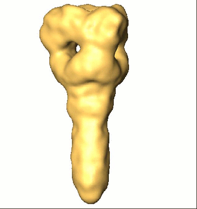 1421.gifemd_1421.tif 1421.gifemd_1421.tif | 43.5 KB 258.8 KB | ||
| Archive directory |  http://ftp.pdbj.org/pub/emdb/structures/EMD-1421ftp://ftp.pdbj.org/pub/emdb/structures/EMD-1421 http://ftp.pdbj.org/pub/emdb/structures/EMD-1421ftp://ftp.pdbj.org/pub/emdb/structures/EMD-1421 | HTTPS FTP |
-Validation report
| Summary document | emd_1421_validation.pdf.gz | 184 KB | Display | EMDB validaton report |
|---|---|---|---|---|
| Full document | emd_1421_full_validation.pdf.gz | 183.1 KB | Display | |
| Data in XML | emd_1421_validation.xml.gz | 5.2 KB | Display | |
| Arichive directory | https://ftp.pdbj.org/pub/emdb/validation_reports/EMD-1421ftp://ftp.pdbj.org/pub/emdb/validation_reports/EMD-1421 | HTTPS FTP |
-Links
| EMDB pages | EMDB (EBI/PDBe) / EMDataResource |
|---|
-Map
| File | Download / File: emd_1421.map.gz / Format: CCP4 / Size: 1.2 MB / Type: IMAGE STORED AS SIGNED BYTE | ||||||||||||||||||||||||||||||||||||||||||||||||||||||||||||||||||||
|---|---|---|---|---|---|---|---|---|---|---|---|---|---|---|---|---|---|---|---|---|---|---|---|---|---|---|---|---|---|---|---|---|---|---|---|---|---|---|---|---|---|---|---|---|---|---|---|---|---|---|---|---|---|---|---|---|---|---|---|---|---|---|---|---|---|---|---|---|---|
| Annotation | In-situ 3D reconstruction of the ectodomain of the PIV5 F protein determined from cryo-negative stain electron micrographs | ||||||||||||||||||||||||||||||||||||||||||||||||||||||||||||||||||||
| Projections & slices | Image control
Images are generated by Spider. | ||||||||||||||||||||||||||||||||||||||||||||||||||||||||||||||||||||
| Voxel size | X=Y=Z: 1.56 Å | ||||||||||||||||||||||||||||||||||||||||||||||||||||||||||||||||||||
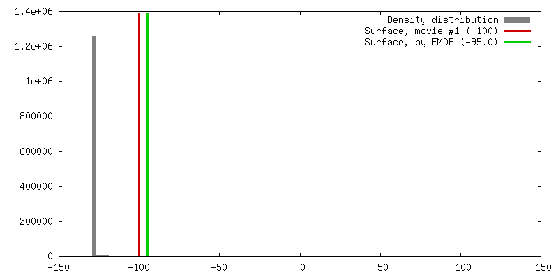
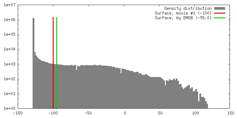
| Density |
| ||||||||||||||||||||||||||||||||||||||||||||||||||||||||||||||||||||
| Symmetry | Space group: 1 | ||||||||||||||||||||||||||||||||||||||||||||||||||||||||||||||||||||
| Details | EMDB XML:
CCP4 map header:
| ||||||||||||||||||||||||||||||||||||||||||||||||||||||||||||||||||||
 Z (Sec.)
 Y (Row.)
 X (Col.)


-Supplemental data
- Sample components
-Entire : F-Protein in intact Parainfluenzavirus 5
| Entire | Name: F-Protein in intact Parainfluenzavirus 5 |
|---|---|
| Components |
|
-Supramolecule #1000: F-Protein in intact Parainfluenzavirus 5
| Supramolecule | Name: F-Protein in intact Parainfluenzavirus 5 / type: sample / ID: 1000 Details: sample of fusion active virions (proven by fluorescence spectroscopy)of PIV5 (strain W3A) Oligomeric state: F-Protein Homotrimer / Number unique components: 1 |
|---|---|
| Molecular weight | Theoretical: 150 KDa |
-Macromolecule #1: F-Protein
| Macromolecule | Name: F-Protein / type: protein_or_peptide / ID: 1 / Oligomeric state: Homotrimer / Recombinant expression: No |
|---|---|
| Source (natural) | Organism: Parainfluenza virus 5 / Strain: W3A / Location in cell: viral membrane |
| Molecular weight | Experimental: 150 KDa |
-Experimental details
-Structure determination
| Method | negative staining, cryo EM |
|---|---|
 Processing Processing | single particle reconstruction |
| Aggregation state | particle |
-Sample preparation
| Buffer | pH: 7.4 / Details: PBS (150 mM NaCl, 5.8 mM NaH2PO4/Na2HPO4) |
|---|---|
| Staining | Type: NEGATIVE Details: 30 seconds absorption 60 seconds staining (1% phospho-tungstic acid, pH 7.4) vitrified in liquid ethane |
| Grid | Details: 200 mesh carbon coated collodium-supported copper grids |
| Vitrification | Cryogen name: ETHANE / Instrument: HOMEMADE PLUNGER / Details: Vitrification instrument: self-construction Method: A small vial of ethane is placed inside a larger liquid nitrogen reservoir. The grid holding a few microliters of the sample is held in place at the bottom of a plunger by the means of fine ...Method: A small vial of ethane is placed inside a larger liquid nitrogen reservoir. The grid holding a few microliters of the sample is held in place at the bottom of a plunger by the means of fine tweezers. Once the ethane in the vial is completely frozen, it needs to be slightly melted. When the liquid ethane is ready, a piece of filter paper is then pressed against the sample to blot of excess buffer, sufficient to leave a thin layer on the grid. After a redetermined time, the filter paper is removed, and the plunger is allowed to drop into the liquid ethane. Once the grid enters the liquid ethane, the sample is rapidly frozen,and the grid is transferred under liquid nitrogen to a storage box immersed liquid nitrogen for later use in the microscope. |
- Electron microscopy
| Microscope | FEI TECNAI F20 |
|---|---|
| Temperature | Average: 92 K |
| Alignment procedure | Legacy - Astigmatism: objective lens astigmatism was corrected at 100,000 times magnification |
| Image recording | Category: FILM / Film or detector model: KODAK SO-163 FILM / Digitization - Scanner: PRIMESCAN / Digitization - Sampling interval: 4 µm / Number real images: 175 / Average electron dose: 12 e/Å2 / Bits/pixel: 8 |
| Electron beam | Acceleration voltage: 160 kV / Electron source:  FIELD EMISSION GUN FIELD EMISSION GUN |
| Electron optics | Calibrated magnification: 51064 / Illumination mode: FLOOD BEAM / Imaging mode: BRIGHT FIELD / Cs: 2 mm / Nominal defocus max: 2.066 µm / Nominal defocus min: 1.501 µm / Nominal magnification: 50000 |
| Sample stage | Specimen holder: Side entry liquid nitrogen-cooled cryo specimen holder Specimen holder model: GATAN LIQUID NITROGEN |
| Experimental equipment |  Model: Tecnai F20 / Image courtesy: FEI Company |
-Image processing
| Details | well resolved spike-like proteins protruding from the viral membrane suitable for single particle analysis were interactively selected |
|---|---|
| CTF correction | Details: MSA-based |
| Final reconstruction | Applied symmetry - Point group: C3 (3 fold cyclic) / Algorithm: OTHER / Resolution.type: BY AUTHOR / Resolution: 15.0 Å / Resolution method: FSC 3 SIGMA CUT-OFF / Software - Name: Imagic / Number images used: 5700 |
