 ムービー
ムービー コントローラー


+ データを開く

- 基本情報
基本情報
| 登録情報 | データベース: EMDB / ID: EMD-1421 | |||||||||
|---|---|---|---|---|---|---|---|---|---|---|
| タイトル | Electron cryomicroscopy reveals different F1+F2 protein States in intact parainfluenza virions. | |||||||||
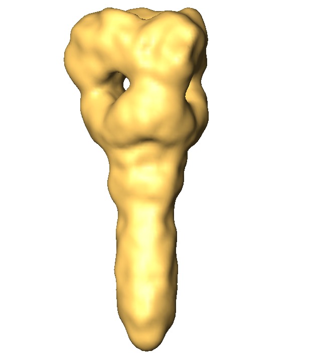
 マップデータ マップデータ | In-situ 3D reconstruction of the ectodomain of the PIV5 F protein determined from cryo-negative stain electron micrographs | |||||||||
 試料 試料 |
| |||||||||
| 生物種 |   Parainfluenza virus 5 (インフルエンザウイルス) Parainfluenza virus 5 (インフルエンザウイルス) | |||||||||
| 手法 | 単粒子再構成法 / クライオ電子顕微鏡法 / ネガティブ染色法 / 解像度: 15.0 Å | |||||||||
 データ登録者 データ登録者 | Ludwig K / Schade B / Boettcher C / Korte T | |||||||||
 引用 引用 | ジャーナル: J Virol / 年: 2008 タイトル: Electron cryomicroscopy reveals different F1+F2 protein States in intact parainfluenza virions. 著者: Kai Ludwig / Boris Schade / Christoph Böttcher / Thomas Korte / Nina Ohlwein / Bolormaa Baljinnyam / Michael Veit / Andreas Herrmann /  要旨: Electron cryomicrographs of intact parainfluenza virus 5 (PIV5) virions revealed two different surface structures, namely, a continuous layer and distinct individual spikes. The structure of these ...Electron cryomicrographs of intact parainfluenza virus 5 (PIV5) virions revealed two different surface structures, namely, a continuous layer and distinct individual spikes. The structure of these spikes reconstructed from intact virions was compared with known F ectodomain structures and was found to be different from the prefusion PIV5 F0 structure but, surprisingly, very similar to the human PIV3 F postfusion structure. Hence, we conclude that the individual F1+F2 spikes in intact PIV5 virions also correspond to the postfusion state. Since the observed fusion activity of PIV5 virions has to be associated with prefusion F1+F2 proteins, they have necessarily to be localized in the continuous surface structure. The data therefore strongly suggest that the prefusion state of the F1+F2 protein requires stabilization, most probably by the association with hemagglutinin-neuraminidase. The conversion of F1+F2 proteins from the prefusion toward the postfusion state while embedded in the virus membrane is topologically difficult to comprehend on the basis of established models and demands reconsideration of our current understanding. | |||||||||
| 履歴 |
|
- 構造の表示
構造の表示
| ムービー |
 ムービービューア ムービービューア |
|---|---|
| 構造ビューア | EMマップ: SurfViewMolmilJmol/JSmol |
| 添付画像 |

- ダウンロードとリンク
-EMDBアーカイブ
| マップデータ | emd_1421.map.gz | 75.5 KB | EMDBマップデータ形式 | |
|---|---|---|---|---|
| ヘッダ (付随情報) | emd-1421-v30.xmlemd-1421.xml | 10.5 KB 10.5 KB | 表示 表示 | EMDBヘッダ |
| 画像 | 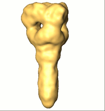 1421.gifemd_1421.tif 1421.gifemd_1421.tif | 43.5 KB 258.8 KB | ||
| アーカイブディレクトリ |  http://ftp.pdbj.org/pub/emdb/structures/EMD-1421ftp://ftp.pdbj.org/pub/emdb/structures/EMD-1421 http://ftp.pdbj.org/pub/emdb/structures/EMD-1421ftp://ftp.pdbj.org/pub/emdb/structures/EMD-1421 | HTTPS FTP |
-リンク
| EMDBのページ | EMDB (EBI/PDBe) / EMDataResource |
|---|
-マップ
| ファイル | ダウンロード / ファイル: emd_1421.map.gz / 形式: CCP4 / 大きさ: 1.2 MB / タイプ: IMAGE STORED AS SIGNED BYTE | ||||||||||||||||||||||||||||||||||||||||||||||||||||||||||||||||||||
|---|---|---|---|---|---|---|---|---|---|---|---|---|---|---|---|---|---|---|---|---|---|---|---|---|---|---|---|---|---|---|---|---|---|---|---|---|---|---|---|---|---|---|---|---|---|---|---|---|---|---|---|---|---|---|---|---|---|---|---|---|---|---|---|---|---|---|---|---|---|
| 注釈 | In-situ 3D reconstruction of the ectodomain of the PIV5 F protein determined from cryo-negative stain electron micrographs | ||||||||||||||||||||||||||||||||||||||||||||||||||||||||||||||||||||
| 投影像・断面図 | 画像のコントロール
画像は Spider により作成 | ||||||||||||||||||||||||||||||||||||||||||||||||||||||||||||||||||||
| ボクセルのサイズ | X=Y=Z: 1.56 Å | ||||||||||||||||||||||||||||||||||||||||||||||||||||||||||||||||||||
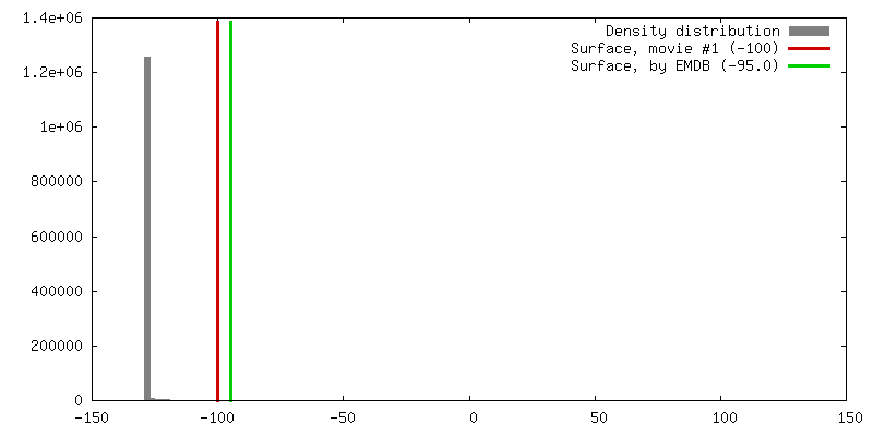
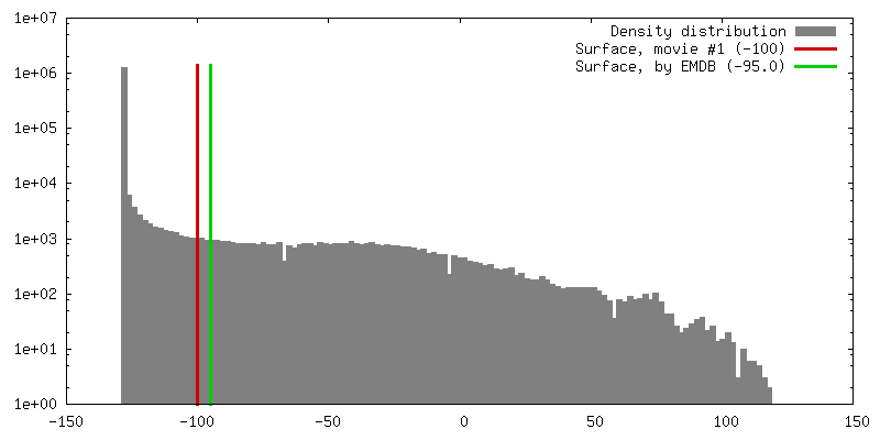
| 密度 |
| ||||||||||||||||||||||||||||||||||||||||||||||||||||||||||||||||||||
| 対称性 | 空間群: 1 | ||||||||||||||||||||||||||||||||||||||||||||||||||||||||||||||||||||
| 詳細 | EMDB XML:
CCP4マップ ヘッダ情報:
| ||||||||||||||||||||||||||||||||||||||||||||||||||||||||||||||||||||
 Z (Sec.)
 Y (Row.)
 X (Col.)


-添付データ
- 試料の構成要素
-全体 : F-Protein in intact Parainfluenzavirus 5
| 全体 | 名称: F-Protein in intact Parainfluenzavirus 5 |
|---|---|
| 要素 |
|
-超分子 #1000: F-Protein in intact Parainfluenzavirus 5
| 超分子 | 名称: F-Protein in intact Parainfluenzavirus 5 / タイプ: sample / ID: 1000 詳細: sample of fusion active virions (proven by fluorescence spectroscopy)of PIV5 (strain W3A) 集合状態: F-Protein Homotrimer / Number unique components: 1 |
|---|---|
| 分子量 | 理論値: 150 KDa |
-分子 #1: F-Protein
| 分子 | 名称: F-Protein / タイプ: protein_or_peptide / ID: 1 / 集合状態: Homotrimer / 組換発現: No |
|---|---|
| 由来(天然) | 生物種: Parainfluenza virus 5 (インフルエンザウイルス) 株: W3A / 細胞中の位置: viral membrane |
| 分子量 | 実験値: 150 KDa |
-実験情報
-構造解析
| 手法 | ネガティブ染色法, クライオ電子顕微鏡法 |
|---|---|
 解析 解析 | 単粒子再構成法 |
| 試料の集合状態 | particle |
-試料調製
| 緩衝液 | pH: 7.4 / 詳細: PBS (150 mM NaCl, 5.8 mM NaH2PO4/Na2HPO4) |
|---|---|
| 染色 | タイプ: NEGATIVE 詳細: 30 seconds absorption 60 seconds staining (1% phospho-tungstic acid, pH 7.4) vitrified in liquid ethane |
| グリッド | 詳細: 200 mesh carbon coated collodium-supported copper grids |
| 凍結 | 凍結剤: ETHANE / 装置: HOMEMADE PLUNGER / 詳細: Vitrification instrument: self-construction 手法: A small vial of ethane is placed inside a larger liquid nitrogen reservoir. The grid holding a few microliters of the sample is held in place at the bottom of a plunger by the means of fine ...手法: A small vial of ethane is placed inside a larger liquid nitrogen reservoir. The grid holding a few microliters of the sample is held in place at the bottom of a plunger by the means of fine tweezers. Once the ethane in the vial is completely frozen, it needs to be slightly melted. When the liquid ethane is ready, a piece of filter paper is then pressed against the sample to blot of excess buffer, sufficient to leave a thin layer on the grid. After a redetermined time, the filter paper is removed, and the plunger is allowed to drop into the liquid ethane. Once the grid enters the liquid ethane, the sample is rapidly frozen,and the grid is transferred under liquid nitrogen to a storage box immersed liquid nitrogen for later use in the microscope. |
- 電子顕微鏡法
| 顕微鏡 | FEI TECNAI F20 |
|---|---|
| 電子線 | 加速電圧: 160 kV / 電子線源: FIELD EMISSION GUN |
| 電子光学系 | 倍率(補正後): 51064 / 照射モード: FLOOD BEAM / 撮影モード: BRIGHT FIELDBright-field microscopy / Cs: 2 mm / 最大 デフォーカス(公称値): 2.066 µm / 最小 デフォーカス(公称値): 1.501 µm / 倍率(公称値): 50000 |
| 試料ステージ | 試料ホルダー: Side entry liquid nitrogen-cooled cryo specimen holder 試料ホルダーモデル: GATAN LIQUID NITROGEN |
| 温度 | 平均: 92 K |
| アライメント法 | Legacy - 非点収差: objective lens astigmatism was corrected at 100,000 times magnification |
| 撮影 | カテゴリ: FILM / フィルム・検出器のモデル: KODAK SO-163 FILM / デジタル化 - スキャナー: PRIMESCAN / デジタル化 - サンプリング間隔: 4 µm / 実像数: 175 / 平均電子線量: 12 e/Å2 / ビット/ピクセル: 8 |
| 実験機器 |  モデル: Tecnai F20 / 画像提供: FEI Company |
-画像解析
| CTF補正 | 詳細: MSA-based |
|---|---|
| 最終 再構成 | 想定した対称性 - 点群: C3 (3回回転対称) / アルゴリズム: OTHER / 解像度のタイプ: BY AUTHOR / 解像度: 15.0 Å / 解像度の算出法: FSC 3 SIGMA CUT-OFF / ソフトウェア - 名称: Imagic / 使用した粒子像数: 5700 |
| 詳細 | well resolved spike-like proteins protruding from the viral membrane suitable for single particle analysis were interactively selected |
