adenine/cytosine mispair binding / MutS complex / mismatch repair complex / regulation of DNA recombination / mismatched DNA binding / DNA binding, bending / ATP-dependent DNA damage sensor activity / mismatch repair / ADP binding / damaged DNA binding ...adenine/cytosine mispair binding / MutS complex / mismatch repair complex / regulation of DNA recombination / mismatched DNA binding / DNA binding, bending / ATP-dependent DNA damage sensor activity / mismatch repair / ADP binding / damaged DNA binding / DNA damage response / ATP hydrolysis activity / ATP binding / identical protein binding / cytosol Similarity search - Function
DNA mismatch repair protein MutS / DNA mismatch repair protein MutS/MSH / DNA mismatch repair protein MutS-like, N-terminal / DNA mismatch repair protein MutS, connector domain / DNA mismatch repair protein MutS, clamp / DNA mismatch repair protein MutS, N-terminal / MutS, connector domain superfamily / MutS domain I / MutS domain II / MutS family domain IV ...DNA mismatch repair protein MutS / DNA mismatch repair protein MutS/MSH / DNA mismatch repair protein MutS-like, N-terminal / DNA mismatch repair protein MutS, connector domain / DNA mismatch repair protein MutS, clamp / DNA mismatch repair protein MutS, N-terminal / MutS, connector domain superfamily / MutS domain I / MutS domain II / MutS family domain IV / MutS domain III / DNA mismatch repair MutS family / DNA mismatch repair protein MutS, C-terminal / DNA mismatch repair protein MutS, core / DNA mismatch repair protein MutS, core domain superfamily / MutS domain V / DNA mismatch repair proteins mutS family signature. / DNA-binding domain of DNA mismatch repair MUTS family / ATPase domain of DNA mismatch repair MUTS family / P-loop containing nucleoside triphosphate hydrolase Similarity search - Domain/homology
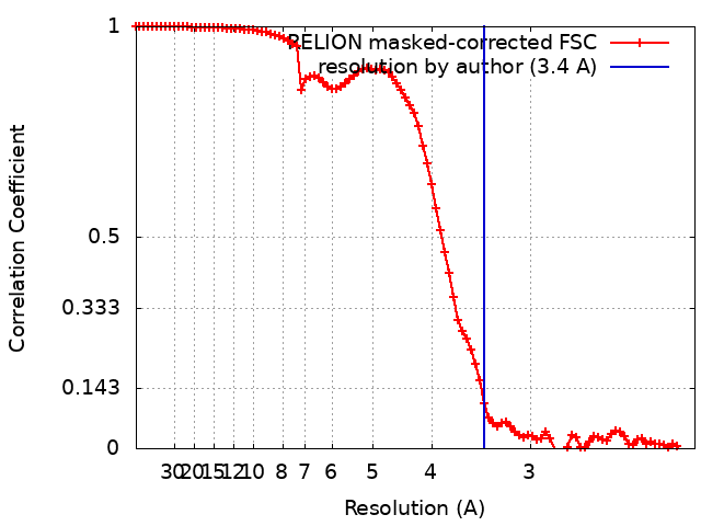
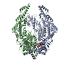
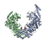
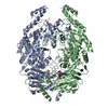
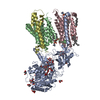
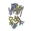
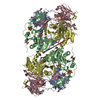
Journal: Nat Struct Mol Biol / Year: 2022 Title: Cryogenic electron microscopy structures reveal how ATP and DNA binding in MutS coordinates sequential steps of DNA mismatch repair. Authors: Alessandro Borsellini / Vladislav Kunetsky / Peter Friedhoff / Meindert H Lamers / Abstract: DNA mismatch repair detects and corrects mismatches introduced during DNA replication. The protein MutS scans for mismatches and coordinates the repair cascade. During this process, MutS undergoes ...DNA mismatch repair detects and corrects mismatches introduced during DNA replication. The protein MutS scans for mismatches and coordinates the repair cascade. During this process, MutS undergoes multiple conformational changes in response to ATP binding, hydrolysis and release, but how ATP induces the various MutS conformations is incompletely understood. Here we present four cryogenic electron microscopy structures of Escherichia coli MutS at sequential stages of the ATP hydrolysis cycle that reveal how ATP binding and hydrolysis induce closing and opening of the MutS dimer, respectively. Biophysical analysis demonstrates how DNA binding modulates the ATPase cycle by prevention of hydrolysis during scanning and mismatch binding, while preventing ADP release in the sliding clamp state. Nucleotide release is achieved when MutS encounters single-stranded DNA that is produced during removal of the daughter strand. The combination of ATP binding and hydrolysis and its modulation by DNA enables MutS to adopt the different conformations needed to coordinate the sequential steps of the mismatch repair cascade.
History
Deposition
Jun 10, 2021
-
Header (metadata) release
Jan 12, 2022
-
Map release
Jan 12, 2022
-
Update
Oct 26, 2022
-
Current status
Oct 26, 2022
Processing site: PDBe / Status: Released
-
Structure visualization
Movie
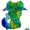
Surface view with section colored by density value
In the structure databanks used in Yorodumi, some data are registered as the other names, "COVID-19 virus" and "2019-nCoV". Here are the details of the virus and the list of structure data.
Jan 31, 2019. EMDB accession codes are about to change! (news from PDBe EMDB page)
EMDB accession codes are about to change! (news from PDBe EMDB page)
The allocation of 4 digits for EMDB accession codes will soon come to an end. Whilst these codes will remain in use, new EMDB accession codes will include an additional digit and will expand incrementally as the available range of codes is exhausted. The current 4-digit format prefixed with “EMD-” (i.e. EMD-XXXX) will advance to a 5-digit format (i.e. EMD-XXXXX), and so on. It is currently estimated that the 4-digit codes will be depleted around Spring 2019, at which point the 5-digit format will come into force.
The EM Navigator/Yorodumi systems omit the EMD- prefix.
Related info.:Q: What is EMD? / ID/Accession-code notation in Yorodumi/EM Navigator
Yorodumi is a browser for structure data from EMDB, PDB, SASBDB, etc.
This page is also the successor to EM Navigator detail page, and also detail information page/front-end page for Omokage search.
The word "yorodu" (or yorozu) is an old Japanese word meaning "ten thousand". "mi" (miru) is to see.
Related info.:EMDB / PDB / SASBDB / Comparison of 3 databanks / Yorodumi Search / Aug 31, 2016. New EM Navigator & Yorodumi / Yorodumi Papers / Jmol/JSmol / Function and homology information / Changes in new EM Navigator and Yorodumi
 Movie
Movie Controller


 Open data
 Basic information
 Map data
 Sample
 Function and homology information
 MutS complex /
 Authors
 Citation

 Structure visualization
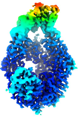
 Downloads & links
 emd_13063.png
 http://ftp.pdbj.org/pub/emdb/structures/EMD-13063


 Sample components
 Processing
 Electron microscopy