Movie
Movie Controller

[English] 日本語
 Yorodumi
- EMDB-1246: Distribution and three-dimensional structure of AIDS virus envelo... -
+ Open data

- Basic information
| Entry | Database: EMDB / ID: EMD-1246 | |||||||||
|---|---|---|---|---|---|---|---|---|---|---|
| Title | Distribution and three-dimensional structure of AIDS virus envelope spikes. | |||||||||
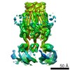
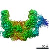
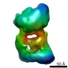
 Map data Map data | 3D map of SIVmac239 envelope spike. It is the 3D averaged volume of all spikes including both "top/bottom view" and "side view" subsets (supplementary method of the reference). Author's threshold 2.65. It should be loaded together with the associated viral membrane map (SIVmac239_Vir_memb.map, EMD-1247, author's threshold 2.25 ). | |||||||||
 Sample Sample |
| |||||||||
| Biological species |  Simian immunodeficiency virus Simian immunodeficiency virus | |||||||||
| Method | subtomogram averaging / cryo EM / Resolution: 32.0 Å | |||||||||
 Authors Authors | Zhu P / Liu J / Bess J / Chertova E / Lifson JD / Grise H / Ofek GA / Taylor KA / Roux KH | |||||||||
 Citation Citation | Journal: Nature / Year: 2006 Title: Distribution and three-dimensional structure of AIDS virus envelope spikes. Authors: Ping Zhu / Jun Liu / Julian Bess / Elena Chertova / Jeffrey D Lifson / Henry Grisé / Gilad A Ofek / Kenneth A Taylor / Kenneth H Roux /  Abstract: Envelope glycoprotein (Env) spikes on AIDS retroviruses initiate infection of host cells and are therefore targets for vaccine development. Though crystal structures for partial Env subunits are ...Envelope glycoprotein (Env) spikes on AIDS retroviruses initiate infection of host cells and are therefore targets for vaccine development. Though crystal structures for partial Env subunits are known, the structure and distribution of native Env spikes on virions is obscure. We applied cryoelectron microscopy tomography to define ultrastructural details of spikes. Virions of wild-type human immunodeficiency virus 1 (HIV-1) and a mutant simian immunodeficiency virus (SIV) had approximately 14 and approximately 73 spikes per particle, respectively, with some clustering of HIV-1 spikes. Three-dimensional averaging showed that the surface glycoprotein (gp120) 'head' of each subunit of the trimeric SIV spike contains a primary mass, with two secondary lobes. The transmembrane glycoprotein 'stalk' of each trimer is composed of three independent legs that project obliquely from the trimer head, tripod-like. Reconciling available atomic structures with the three-dimensional whole spike density map yields insights into the orientation of Env spike structural elements and possible structural bases of their functions. | |||||||||
| History |
|
- Structure visualization
| Movie |
 Movie viewer Movie viewer |
|---|---|
| Structure viewer | EM map: SurfViewMolmilJmol/JSmol |
| Supplemental images |
- Downloads & links
-EMDB archive
| Map data | emd_1246.map.gz | 1.3 MB | EMDB map data format | |
|---|---|---|---|---|
| Header (meta data) | emd-1246-v30.xmlemd-1246.xml | 11.3 KB 11.3 KB | Display Display | EMDB header |
| Images |  1246.gif 1246.gif | 57.6 KB | ||
| Archive directory |  http://ftp.pdbj.org/pub/emdb/structures/EMD-1246ftp://ftp.pdbj.org/pub/emdb/structures/EMD-1246 http://ftp.pdbj.org/pub/emdb/structures/EMD-1246ftp://ftp.pdbj.org/pub/emdb/structures/EMD-1246 | HTTPS FTP |
-Validation report
| Summary document | emd_1246_validation.pdf.gz | 201.2 KB | Display | EMDB validaton report |
|---|---|---|---|---|
| Full document | emd_1246_full_validation.pdf.gz | 200.3 KB | Display | |
| Data in XML | emd_1246_validation.xml.gz | 5.3 KB | Display | |
| Arichive directory | https://ftp.pdbj.org/pub/emdb/validation_reports/EMD-1246ftp://ftp.pdbj.org/pub/emdb/validation_reports/EMD-1246 | HTTPS FTP |
-Related structure data


-Links
| EMDB pages | EMDB (EBI/PDBe) / EMDataResource |
|---|
-Map
| File | Download / File: emd_1246.map.gz / Format: CCP4 / Size: 1.4 MB / Type: IMAGE STORED AS FLOATING POINT NUMBER (4 BYTES) | ||||||||||||||||||||||||||||||||||||||||||||||||||||||||||||||||||||
|---|---|---|---|---|---|---|---|---|---|---|---|---|---|---|---|---|---|---|---|---|---|---|---|---|---|---|---|---|---|---|---|---|---|---|---|---|---|---|---|---|---|---|---|---|---|---|---|---|---|---|---|---|---|---|---|---|---|---|---|---|---|---|---|---|---|---|---|---|---|
| Annotation | 3D map of SIVmac239 envelope spike. It is the 3D averaged volume of all spikes including both "top/bottom view" and "side view" subsets (supplementary method of the reference). Author's threshold 2.65. It should be loaded together with the associated viral membrane map (SIVmac239_Vir_memb.map, EMD-1247, author's threshold 2.25 ). | ||||||||||||||||||||||||||||||||||||||||||||||||||||||||||||||||||||
| Voxel size | X=Y=Z: 5.56 Å | ||||||||||||||||||||||||||||||||||||||||||||||||||||||||||||||||||||
| Density |
| ||||||||||||||||||||||||||||||||||||||||||||||||||||||||||||||||||||
| Symmetry | Space group: 1 | ||||||||||||||||||||||||||||||||||||||||||||||||||||||||||||||||||||
| Details | EMDB XML:
CCP4 map header:
| ||||||||||||||||||||||||||||||||||||||||||||||||||||||||||||||||||||
-Supplemental data
- Sample components
-Entire : Envelope Spike on the surface of SIVmac239 virus with truncated c...
| Entire | Name: Envelope Spike on the surface of SIVmac239 virus with truncated cytoplasmic tail |
|---|---|
| Components |
|
-Supramolecule #1000: Envelope Spike on the surface of SIVmac239 virus with truncated c...
| Supramolecule | Name: Envelope Spike on the surface of SIVmac239 virus with truncated cytoplasmic tail type: sample / ID: 1000 Details: The virus was AT-2 treated to eliminate the infectivity. Oligomeric state: Trimer / Number unique components: 1 |
|---|---|
| Molecular weight | Theoretical: 450 KDa |
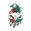
-Macromolecule #1: Envelope glycoprotein
| Macromolecule | Name: Envelope glycoprotein / type: protein_or_peptide / ID: 1 / Name.synonym: gp120, gp41 Details: The spike consists of 3 copies of gp120 and gp41 ecto domain to form a trimer Oligomeric state: trimer / Recombinant expression: No / Database: NCBI |
|---|---|
| Source (natural) | Organism: Simian immunodeficiency virus |
| Molecular weight | Experimental: 450 KDa |
-Experimental details
-Structure determination
| Method | cryo EM |
|---|---|
 Processing Processing | subtomogram averaging |
-Sample preparation
| Buffer | Details: PBS |
|---|---|
| Grid | Details: 200 mesh Quantifoil R2/1 copper grid |
| Vitrification | Cryogen name: ETHANE / Instrument: HOMEMADE PLUNGER / Details: Vitrification instrument: Home made plunger |
- Electron microscopy
| Microscope | FEI/PHILIPS CM300FEG/ST |
|---|---|
| Details | Minimum tilt angle was -70 |
| Date | Aug 30, 2004 |
| Image recording | Category: CCD / Film or detector model: TVIPS TEMCAM-F224 (2k x 2k) / Number real images: 80 / Average electron dose: 60 e/Å2 |
| Electron beam | Acceleration voltage: 300 kV / Electron source:  FIELD EMISSION GUN FIELD EMISSION GUN |
| Electron optics | Calibrated magnification: 43200 / Illumination mode: FLOOD BEAM / Imaging mode: BRIGHT FIELD / Nominal defocus max: 6.0 µm / Nominal defocus min: 4.0 µm |
| Sample stage | Specimen holder: Gatan 626 cryoholder / Specimen holder model: GATAN LIQUID NITROGEN / Tilt series - Axis1 - Min angle: 70 ° / Tilt series - Axis1 - Max angle: 70 ° |
-Image processing
| Details | The tomographic tilt angle increment was determined by cosine rule, i.e., ~2-3 degrees increment around low tilt angles and <1 degree increment in the high tilt angles. Average number of tilts used in the 3D reconstructions: 80. Average tomographic tilt angle increment: 2. |
|---|---|
| Final reconstruction | Algorithm: OTHER / Resolution.type: BY AUTHOR / Resolution: 32.0 Å / Resolution method: OTHER / Software - Name: Protomo Details: The resolution of the final averaged map, determined by Fourier shell correlation (based on a cutoff value of 0.5) is 2.5 nm and, after low pass filtering to the estimated first node in the ...Details: The resolution of the final averaged map, determined by Fourier shell correlation (based on a cutoff value of 0.5) is 2.5 nm and, after low pass filtering to the estimated first node in the contrast transfer function, 3.2 nm. |
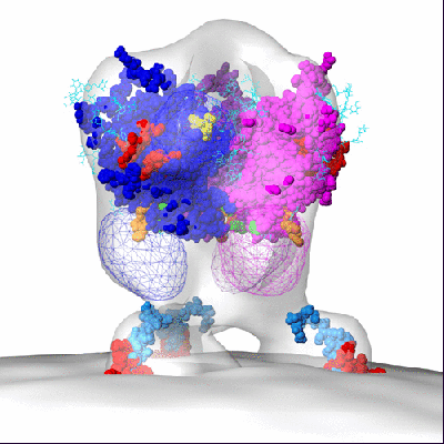
-Atomic model buiding 1
| Initial model |
| ||||||||
|---|---|---|---|---|---|---|---|---|---|
| Software | Name: Chimera | ||||||||
| Details | PDBEntryID_givenInChain. The gp120 core and 2F5 and 4E10 peptides structure were fittted by manual docking using program chimera and then applied 3-fold symmetry around Z axis. | ||||||||
| Refinement | Protocol: RIGID BODY FIT |