 Movie
Movie Controller


+ Open data

- Basic information
| Entry | Database: EMDB / ID: EMD-0650 | ||||||||||||||||||
|---|---|---|---|---|---|---|---|---|---|---|---|---|---|---|---|---|---|---|---|
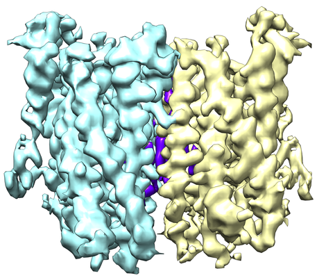
| Title | Cryo-EM structure of OTOP3 from xenopus tropicalis | ||||||||||||||||||
 Map data Map data | Cryo-EM structure of OTOP3 from xenopus tropicalis | ||||||||||||||||||
 Sample Sample |
| ||||||||||||||||||
 Keywords Keywords |  Otopetrin / proton / channel / MEMBRANE PROTEIN Otopetrin / proton / channel / MEMBRANE PROTEIN | ||||||||||||||||||
| Function / homology | Otopetrin / Otopetrin / monoatomic ion transport / membrane => GO:0016020 / plasma membrane / LOC100127796 protein Function and homology information Function and homology information | ||||||||||||||||||
| Biological species | Xenopus tropicalis (tropical clawed frog) | ||||||||||||||||||
| Method | single particle reconstruction / cryo EM / Resolution: 3.92 Å | ||||||||||||||||||
 Authors Authors | Chen QF / Bai XC | ||||||||||||||||||
| Funding support |  United States, 5 items United States, 5 items
| ||||||||||||||||||
 Citation Citation | Journal: Elife / Year: 2019 Title: Structural and functional characterization of an otopetrin family proton channel. Authors: Qingfeng Chen / Weizhong Zeng / Ji She / Xiao-Chen Bai / Youxing Jiang / Abstract: The otopetrin (OTOP) proteins were recently characterized as proton channels. Here we present the cryo-EM structure of OTOP3 from (XtOTOP3) along with functional characterization of the channel. ...The otopetrin (OTOP) proteins were recently characterized as proton channels. Here we present the cryo-EM structure of OTOP3 from (XtOTOP3) along with functional characterization of the channel. XtOTOP3 forms a homodimer with each subunit containing 12 transmembrane helices that can be divided into two structurally homologous halves; each half assembles as an α-helical barrel that could potentially serve as a proton conduction pore. Both pores open from the extracellular half before becoming occluded at a central constriction point consisting of three highly conserved residues - Gln-Asp/Asn-Tyr (the constriction triads). Mutagenesis shows that the constriction triad from the second pore is less amenable to perturbation than that of the first pore, suggesting an unequal contribution between the two pores to proton transport. We also identified several key residues at the interface between the two pores that are functionally important, particularly Asp509, which confers intracellular pH-dependent desensitization to OTOP channels. | ||||||||||||||||||
| History |
|
- Structure visualization
| Movie |
Movie viewer |
|---|---|
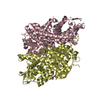
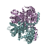
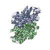
| Structure viewer | EM map: SurfViewMolmilJmol/JSmol |
| Supplemental images |
- Downloads & links
-EMDB archive
| Map data | emd_0650.map.gz | 20.2 MB | EMDB map data format | |
|---|---|---|---|---|
| Header (meta data) | emd-0650-v30.xmlemd-0650.xml | 14.5 KB 14.5 KB | Display Display | EMDB header |
| Images |  emd_0650.png emd_0650.png | 226.6 KB | ||
| Filedesc metadata | emd-0650.cif.gz | 6.3 KB | ||
| Archive directory |  http://ftp.pdbj.org/pub/emdb/structures/EMD-0650ftp://ftp.pdbj.org/pub/emdb/structures/EMD-0650 http://ftp.pdbj.org/pub/emdb/structures/EMD-0650ftp://ftp.pdbj.org/pub/emdb/structures/EMD-0650 | HTTPS FTP |
-Related structure data
| Related structure data |  6o84MC M: atomic model generated by this map C: citing same article ( |
|---|---|
| Similar structure data |


-Links
| EMDB pages | EMDB (EBI/PDBe) / EMDataResource |
|---|
-Map
| File | Download / File: emd_0650.map.gz / Format: CCP4 / Size: 22.2 MB / Type: IMAGE STORED AS FLOATING POINT NUMBER (4 BYTES) | ||||||||||||||||||||||||||||||||||||||||||||||||||||||||||||
|---|---|---|---|---|---|---|---|---|---|---|---|---|---|---|---|---|---|---|---|---|---|---|---|---|---|---|---|---|---|---|---|---|---|---|---|---|---|---|---|---|---|---|---|---|---|---|---|---|---|---|---|---|---|---|---|---|---|---|---|---|---|
| Annotation | Cryo-EM structure of OTOP3 from xenopus tropicalis | ||||||||||||||||||||||||||||||||||||||||||||||||||||||||||||
| Voxel size | X=Y=Z: 1.07 Å | ||||||||||||||||||||||||||||||||||||||||||||||||||||||||||||
| Density |
| ||||||||||||||||||||||||||||||||||||||||||||||||||||||||||||
| Symmetry | Space group: 1 | ||||||||||||||||||||||||||||||||||||||||||||||||||||||||||||
| Details | EMDB XML:
CCP4 map header:
| ||||||||||||||||||||||||||||||||||||||||||||||||||||||||||||
-Supplemental data
- Sample components
-Entire : Xenopus tropicalis OTOP3
| Entire | Name: Xenopus tropicalis OTOP3 |
|---|---|
| Components |
|
-Supramolecule #1: Xenopus tropicalis OTOP3
| Supramolecule | Name: Xenopus tropicalis OTOP3 / type: organelle_or_cellular_component / ID: 1 / Parent: 0 / Macromolecule list: all |
|---|---|
| Source (natural) | Organism: Xenopus tropicalis (tropical clawed frog) |
| Molecular weight | Theoretical: 77 KDa |
-Macromolecule #1: LOC100127796 protein,LOC100127796 protein,OTOP3,LOC100127796 prot...
| Macromolecule | Name: LOC100127796 protein,LOC100127796 protein,OTOP3,LOC100127796 protein,LOC100127796 protein type: protein_or_peptide / ID: 1 / Number of copies: 2 / Enantiomer: LEVO |
|---|---|
| Source (natural) | Organism: Xenopus tropicalis (tropical clawed frog) |
| Molecular weight | Theoretical: 73.352297 KDa |
| Recombinant expression | Organism:  Homo sapiens (human) Homo sapiens (human) |
| Sequence | String: MLSKEEPACR QFHSREKTWG NEHNGKTVTQ ANNKEHKVPA VRRGDIGQRE PAAHPASHIQ GTMASENTGE QATCKDQYMD LDAPGNNLE HSWLHRHCEI PTTLHQRAKK TGRLFSGLFG LNLMFLGGTV VSSVALSNKA VPERDSQSFL CILMLLSSVW A LYHLLFIR ...String: MLSKEEPACR QFHSREKTWG NEHNGKTVTQ ANNKEHKVPA VRRGDIGQRE PAAHPASHIQ GTMASENTGE QATCKDQYMD LDAPGNNLE HSWLHRHCEI PTTLHQRAKK TGRLFSGLFG LNLMFLGGTV VSSVALSNKA VPERDSQSFL CILMLLSSVW A LYHLLFIR NQNGAVHHDH HAGAMWLKAS LAIFGVCSII LSIFEIGHAL LLQNCEILMD IVFFSIEIVF VSVQTVLLWV SC KDCVQMH HSVTRYGIML TLATDILLWL TAVIDDSLEQ DLEILQSNST QDESNEMAQC QCPTDSMCWG LKQGYVTMFP FNI EYSLIC ATLLFIMWKN VGRRE(UNK)(UNK)(UNK)(UNK)(UNK) (UNK)(UNK)(UNK)(UNK)(UNK)(UNK) (UNK)(UNK)(UNK) (UNK)(UNK)(UNK)(UNK)(UNK)(UNK)(UNK)(UNK)(UNK)(UNK) (UNK)(UNK)(UNK) (UNK)(UNK)(UNK)(UNK)(UNK)(UNK) (UNK)(UNK)(UNK)(UNK)(UNK)(UNK)(UNK)(UNK)(UNK)(UNK) (UNK)(UNK)(UNK)(UNK)(UNK)(UNK)(UNK)(UNK)(UNK) (UNK)(UNK)(UNK)(UNK)(UNK)(UNK)(UNK) (UNK)(UNK) (UNK)(UNK)(UNK)(UNK)(UNK)(UNK)(UNK)(UNK)(UNK)(UNK) (UNK)(UNK)(UNK)(UNK) (UNK)(UNK)(UNK)(UNK)(UNK) (UNK)(UNK)(UNK)(UNK)(UNK)(UNK)(UNK)(UNK)(UNK)(UNK) (UNK) (UNK)(UNK)(UNK)(UNK)W(UNK)(UNK)(UNK) (UNK)(UNK)(UNK)(UNK)(UNK)(UNK)(UNK)(UNK) (UNK) (UNK)(UNK)(UNK)(UNK)(UNK)(UNK)(UNK)(UNK)(UNK)(UNK) (UNK)(UNK)(UNK)(UNK)(UNK) (UNK)(UNK)(UNK)(UNK) (UNK)(UNK)(UNK)(UNK)(UNK)(UNK)(UNK)(UNK)(UNK)(UNK) (UNK)(UNK) (UNK)(UNK)(UNK)(UNK)(UNK)(UNK)(UNK) (UNK)(UNK)(UNK)(UNK)(UNK)(UNK)(UNK)(UNK)(UNK) (UNK)(UNK)(UNK)(UNK)(UNK)(UNK)(UNK)(UNK)(UNK)R KLDVTLLFVS AVGQLGISYF SIIATVVTTP WTML SALNF SNSLLLILQY LSQTMFIIES MRSIHEEEKE KPGHHEESHR RMSVQEMHKA PPSCLDAGHL GLSRRVVKEM AMFLM ICNI MCWILGAFGA HPLYMNGLER QLYGSGIWLA ILNIGLPLSV FYRMHSVGIL LEVYLHALEG SSLVPR UniProtKB: LOC100127796 protein, LOC100127796 protein |
-Experimental details
-Structure determination
| Method | cryo EM |
|---|---|
 Processing Processing | single particle reconstruction |
| Aggregation state | particle |
-Sample preparation
| Concentration | 1.3 mg/mL |
|---|---|
| Buffer | pH: 8 / Component - Concentration: 150.0 mM / Component - Formula: NaClSodium chloride / Component - Name: sodium chloride Details: Solutions were made fresh from stock solutions and filtered. |
| Vitrification | Cryogen name: ETHANE / Chamber humidity: 100 % / Chamber temperature: 277 K / Instrument: FEI VITROBOT MARK IV |
| Details | This sample was monodisperse |
- Electron microscopy
| Microscope | FEI TITAN KRIOS |
|---|---|
| Electron beam | Acceleration voltage: 300 kV / Electron source: FIELD EMISSION GUN |
| Electron optics | C2 aperture diameter: 70.0 µm / Illumination mode: FLOOD BEAM / Imaging mode: BRIGHT FIELDBright-field microscopy / Cs: 2.7 mm / Nominal defocus max: 3.0 µm / Nominal defocus min: 1.2 µm / Nominal magnification: 46730 |
| Specialist optics | Energy filter - Name: GIF Quantum LS |
| Sample stage | Specimen holder model: FEI TITAN KRIOS AUTOGRID HOLDER / Cooling holder cryogen: NITROGEN |
| Image recording | Film or detector model: GATAN K2 SUMMIT (4k x 4k) / Detector mode: SUPER-RESOLUTION / Number grids imaged: 1 / Number real images: 3000 / Average exposure time: 15.0 sec. / Average electron dose: 50.0 e/Å2 |
| Experimental equipment |  Model: Titan Krios / Image courtesy: FEI Company |
-Image processing
| Particle selection | Number selected: 1433949 / Details: The particles were auto-picked in RELION. |
|---|---|
| Startup model | Type of model: NONE |
| Initial angle assignment | Type: OTHER / Software - Name: RELION (ver. 2) |
| Final 3D classification | Number classes: 8 / Software - Name: RELION (ver. 2) |
| Final angle assignment | Type: PROJECTION MATCHING / Software - Name: RELION (ver. 2) Details: Auto-refinement in RELION generated the final reconstruction. The angular sampling was determined automatically in RELION. |
| Final reconstruction | Resolution.type: BY AUTHOR / Resolution: 3.92 Å / Resolution method: FSC 0.143 CUT-OFF / Software - Name: RELION (ver. 2) / Number images used: 212551 |
| Details | 30 frames per movie stack were saved for motion correction. |
