3TAM
 
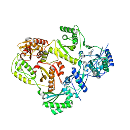 | |
3TJ0
 
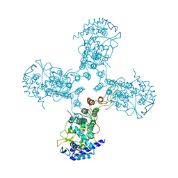 | | Crystal Structure of Influenza B Virus Nucleoprotein | | 分子名称: | Nucleoprotein | | 著者 | Ng, A.K.L, Zhang, H, Liu, J, Au, S.W.N, Wang, J, Shaw, P.C. | | 登録日 | 2011-08-23 | | 公開日 | 2012-06-20 | | 最終更新日 | 2023-11-01 | | 実験手法 | X-RAY DIFFRACTION (3.233 Å) | | 主引用文献 | Structural basis for RNA binding and homo-oligomer formation by influenza B virus nucleoprotein
J.Virol., 86, 2012
|
|
1DV2
 
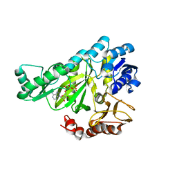 | | The structure of biotin carboxylase, mutant E288K, complexed with ATP | | 分子名称: | ADENOSINE-5'-TRIPHOSPHATE, BIOTIN CARBOXYLASE | | 著者 | Thoden, J.B, Blanchard, C.Z, Holden, H.M, Waldrop, G.L. | | 登録日 | 2000-01-19 | | 公開日 | 2000-06-09 | | 最終更新日 | 2024-05-22 | | 実験手法 | X-RAY DIFFRACTION (2.5 Å) | | 主引用文献 | Movement of the biotin carboxylase B-domain as a result of ATP binding.
J.Biol.Chem., 275, 2000
|
|
5SEY
 
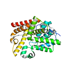 | | Crystal Structure of human phosphodiesterase 10 in complex with [2-cyclopropyl-6-[2-(1-methyl-4-phenylimidazol-2-yl)ethynyl]imidazo[1,2-b]pyridazin-3-yl]methanol | | 分子名称: | MAGNESIUM ION, ZINC ION, cAMP and cAMP-inhibited cGMP 3',5'-cyclic phosphodiesterase 10A, ... | | 著者 | Joseph, C, Groebke-Zbinden, K, Benz, J, Schlatter, D, Rudolph, M.G. | | 登録日 | 2022-01-21 | | 公開日 | 2022-10-12 | | 最終更新日 | 2024-04-03 | | 実験手法 | X-RAY DIFFRACTION (2.29 Å) | | 主引用文献 | A high quality, industrial data set for binding affinity prediction: performance comparison in different early drug discovery scenarios.
J.Comput.Aided Mol.Des., 36, 2022
|
|
5SF5
 
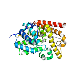 | | Crystal Structure of human phosphodiesterase 10 in complex with 2,3-dimethyl-6-[2-(1-methyl-4-phenylimidazol-2-yl)ethyl]imidazo[1,2-b]pyridazine | | 分子名称: | (4S)-2,3-dimethyl-6-[2-(1-methyl-4-phenyl-1H-imidazol-2-yl)ethyl]imidazo[1,2-b]pyridazine, MAGNESIUM ION, ZINC ION, ... | | 著者 | Joseph, C, Groebke-Zbinden, K, Benz, J, Schlatter, D, Rudolph, M.G. | | 登録日 | 2022-01-21 | | 公開日 | 2022-10-12 | | 最終更新日 | 2024-04-03 | | 実験手法 | X-RAY DIFFRACTION (1.98 Å) | | 主引用文献 | A high quality, industrial data set for binding affinity prediction: performance comparison in different early drug discovery scenarios.
J.Comput.Aided Mol.Des., 36, 2022
|
|
5SFB
 
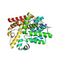 | | Crystal Structure of human phosphodiesterase 10 in complex with N,2,3-trimethyl-6-[2-(2-methyl-5-pyrrolidin-1-yl-1,2,4-triazol-3-yl)ethyl]imidazo[1,2-b]pyridazine-8-carboxamide | | 分子名称: | (4S)-N,2,3-trimethyl-6-{2-[1-methyl-3-(pyrrolidin-1-yl)-1H-1,2,4-triazol-5-yl]ethyl}imidazo[1,2-b]pyridazine-8-carboxamide, MAGNESIUM ION, ZINC ION, ... | | 著者 | Joseph, C, Groebke-Zbinden, K, Benz, J, Schlatter, D, Rudolph, M.G. | | 登録日 | 2022-01-21 | | 公開日 | 2022-10-12 | | 最終更新日 | 2024-04-03 | | 実験手法 | X-RAY DIFFRACTION (1.98 Å) | | 主引用文献 | A high quality, industrial data set for binding affinity prediction: performance comparison in different early drug discovery scenarios.
J.Comput.Aided Mol.Des., 36, 2022
|
|
5SFF
 
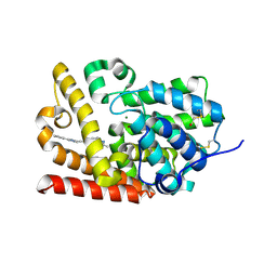 | | Crystal Structure of human phosphodiesterase 10 in complex with [2-methyl-6-[2-(1-methyl-4-phenylimidazol-2-yl)ethynyl]imidazo[1,2-b]pyridazin-3-yl]methanol | | 分子名称: | MAGNESIUM ION, ZINC ION, cAMP and cAMP-inhibited cGMP 3',5'-cyclic phosphodiesterase 10A, ... | | 著者 | Joseph, C, Groebke-Zbinden, K, Benz, J, Schlatter, D, Rudolph, M.G. | | 登録日 | 2022-01-21 | | 公開日 | 2022-10-12 | | 最終更新日 | 2024-04-03 | | 実験手法 | X-RAY DIFFRACTION (2.16 Å) | | 主引用文献 | A high quality, industrial data set for binding affinity prediction: performance comparison in different early drug discovery scenarios.
J.Comput.Aided Mol.Des., 36, 2022
|
|
2QC5
 
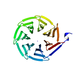 | | Streptogramin B lyase structure | | 分子名称: | IODIDE ION, Streptogramin B lactonase | | 著者 | Lipka, M, Bochtler, M. | | 登録日 | 2007-06-19 | | 公開日 | 2008-10-14 | | 最終更新日 | 2024-02-21 | | 実験手法 | X-RAY DIFFRACTION (1.8 Å) | | 主引用文献 | Crystal structure and mechanism of the Staphylococcus cohnii virginiamycin B lyase (Vgb).
Biochemistry, 47, 2008
|
|
1SQB
 
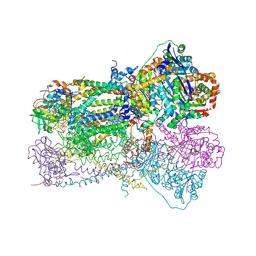 | | Crystal Structure Analysis of Bovine Bc1 with Azoxystrobin | | 分子名称: | Cytochrome b, Cytochrome c1, heme protein, ... | | 著者 | Esser, L, Quinn, B, Li, Y.F, Zhang, M, Elberry, M, Yu, L, Yu, C.A, Xia, D. | | 登録日 | 2004-03-18 | | 公開日 | 2004-09-07 | | 最終更新日 | 2023-08-23 | | 実験手法 | X-RAY DIFFRACTION (2.69 Å) | | 主引用文献 | Crystallographic studies of quinol oxidation site inhibitors: a modified classification of inhibitors for the cytochrome bc(1) complex
J.Mol.Biol., 341, 2004
|
|
5SHL
 
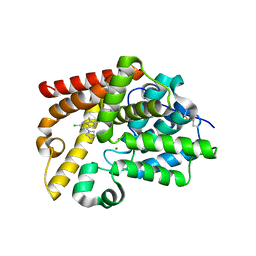 | | Crystal Structure of human phosphodiesterase 10 in complex with 6-chloro-2,3-dimethyl-8-pyrrolidin-1-ylimidazo[1,2-b]pyridazine | | 分子名称: | (4R)-6-chloro-2,3-dimethyl-8-(pyrrolidin-1-yl)imidazo[1,2-b]pyridazine, MAGNESIUM ION, ZINC ION, ... | | 著者 | Joseph, C, Benz, J, Flohr, A, Groebke-Zbinden, K, Rudolph, M.G. | | 登録日 | 2022-02-01 | | 公開日 | 2022-10-12 | | 最終更新日 | 2024-04-03 | | 実験手法 | X-RAY DIFFRACTION (2.35 Å) | | 主引用文献 | Crystal Structure of a human phosphodiesterase 10 complex
To be published
|
|
5SJK
 
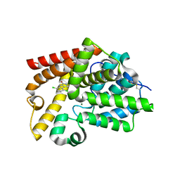 | | Crystal Structure of human phosphodiesterase 10 in complex with 5,8-dichloro-2,3,4,9-tetrahydropyrido[3,4-b]indol-1-one | | 分子名称: | 5,8-dichloro-2,3,4,9-tetrahydro-1H-pyrido[3,4-b]indol-1-one, MAGNESIUM ION, ZINC ION, ... | | 著者 | Joseph, C, Benz, J, Flohr, A, Kyburz, E, Rudolph, M.G. | | 登録日 | 2022-02-01 | | 公開日 | 2022-10-12 | | 最終更新日 | 2024-04-03 | | 実験手法 | X-RAY DIFFRACTION (2.24 Å) | | 主引用文献 | Crystal Structure of a human phosphodiesterase 10 complex
To be published
|
|
1CZ1
 
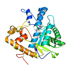 | | EXO-B-(1,3)-GLUCANASE FROM CANDIDA ALBICANS AT 1.85 A RESOLUTION | | 分子名称: | PROTEIN (EXO-B-(1,3)-GLUCANASE) | | 著者 | Cutfield, S.M, Davies, G.J, Murshudov, G, Anderson, B.F, Moody, P.C.E, Sullivan, P.A, Cutfield, J.F. | | 登録日 | 1999-09-01 | | 公開日 | 2000-01-03 | | 最終更新日 | 2017-10-04 | | 実験手法 | X-RAY DIFFRACTION (1.85 Å) | | 主引用文献 | The structure of the exo-beta-(1,3)-glucanase from Candida albicans in native and bound forms: relationship between a pocket and groove in family 5 glycosyl hydrolases.
J.Mol.Biol., 294, 1999
|
|
4O8U
 
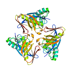 | | Structure of PF2046 | | 分子名称: | Uncharacterized protein PF2046 | | 著者 | Su, J, Liu, Z.-J. | | 登録日 | 2013-12-30 | | 公開日 | 2014-04-30 | | 実験手法 | X-RAY DIFFRACTION (2.345 Å) | | 主引用文献 | Crystal structure of a novel non-Pfam protein PF2046 solved using low resolution B-factor sharpening and multi-crystal averaging methods
Protein Cell, 1, 2010
|
|
5SF1
 
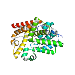 | | Crystal Structure of human phosphodiesterase 10 in complex with 2-(difluoromethyl)-3-methyl-6-[2-(1-methyl-4-phenylimidazol-2-yl)ethynyl]imidazo[1,2-b]pyridazine | | 分子名称: | (4S)-2-(difluoromethyl)-3-methyl-6-[(1-methyl-4-phenyl-1H-imidazol-2-yl)ethynyl]imidazo[1,2-b]pyridazine, MAGNESIUM ION, ZINC ION, ... | | 著者 | Joseph, C, Groebke-Zbinden, K, Benz, J, Schlatter, D, Rudolph, M.G. | | 登録日 | 2022-01-21 | | 公開日 | 2022-10-12 | | 最終更新日 | 2024-04-03 | | 実験手法 | X-RAY DIFFRACTION (2.11 Å) | | 主引用文献 | A high quality, industrial data set for binding affinity prediction: performance comparison in different early drug discovery scenarios.
J.Comput.Aided Mol.Des., 36, 2022
|
|
5SIE
 
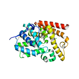 | | Crystal Structure of human phosphodiesterase 10 in complex with 5-chloro-8-hydroxy-2-methyl-1,4-dihydropyrrolo[3,4-b]indol-3-one | | 分子名称: | 5-chloro-8-hydroxy-2-methyl-1,4-dihydropyrrolo[3,4-b]indol-3(2H)-one, MAGNESIUM ION, ZINC ION, ... | | 著者 | Joseph, C, Benz, J, Flohr, A, Boehringer, M, Rudolph, M.G. | | 登録日 | 2022-02-01 | | 公開日 | 2022-10-12 | | 最終更新日 | 2024-04-03 | | 実験手法 | X-RAY DIFFRACTION (2.12 Å) | | 主引用文献 | Crystal Structure of a human phosphodiesterase 10 complex
To be published
|
|
5SEM
 
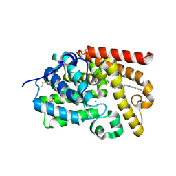 | | Crystal Structure of human phosphodiesterase 10 in complex with 3-methyl-6-[2-(1-methyl-4-phenylimidazol-2-yl)ethyl]-2-(trifluoromethyl)imidazo[1,2-b]pyridazine | | 分子名称: | (4S)-3-methyl-6-[2-(1-methyl-4-phenyl-1H-imidazol-2-yl)ethyl]-2-(trifluoromethyl)imidazo[1,2-b]pyridazine, MAGNESIUM ION, ZINC ION, ... | | 著者 | Joseph, C, Groebke-Zbinden, K, Benz, J, Schlatter, D, Rudolph, M.G. | | 登録日 | 2022-01-21 | | 公開日 | 2022-10-12 | | 最終更新日 | 2024-04-03 | | 実験手法 | X-RAY DIFFRACTION (2.1 Å) | | 主引用文献 | A high quality, industrial data set for binding affinity prediction: performance comparison in different early drug discovery scenarios.
J.Comput.Aided Mol.Des., 36, 2022
|
|
5SKB
 
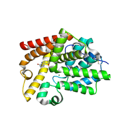 | | Crystal Structure of human phosphodiesterase 10 in complex with 9-methoxy-3,5-dihydropyrimido[5,4-b]indol-4-one | | 分子名称: | 9-methoxy-3,5-dihydro-4H-pyrimido[5,4-b]indol-4-one, MAGNESIUM ION, ZINC ION, ... | | 著者 | Joseph, C, Benz, J, Flohr, A, Brunner, M, Rudolph, M.G. | | 登録日 | 2022-02-01 | | 公開日 | 2022-10-12 | | 最終更新日 | 2024-04-03 | | 実験手法 | X-RAY DIFFRACTION (2.35 Å) | | 主引用文献 | Crystal Structure of a human phosphodiesterase 10 complex
To be published
|
|
4OLR
 
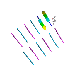 | | [Leu-5]-Enkephalin mutant - YVVFV | | 分子名称: | (4S)-2-METHYL-2,4-PENTANEDIOL, [Leu-5]-Enkephalin mutant - YVVFV | | 著者 | Sangwan, S, Eisenberg, D, Sawaya, M.R, Do, T.D, Bowers, M.T, Lapointe, N.E, Teplow, D.B, Feinstein, S.C. | | 登録日 | 2014-01-24 | | 公開日 | 2014-07-02 | | 最終更新日 | 2024-02-28 | | 実験手法 | X-RAY DIFFRACTION (1.1 Å) | | 主引用文献 | Factors that drive Peptide assembly from native to amyloid structures: experimental and theoretical analysis of [leu-5]-enkephalin mutants.
J.Phys.Chem.B, 118, 2014
|
|
5SK3
 
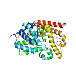 | | Crystal Structure of human phosphodiesterase 10 in complex with [2-cyclopropyl-6-[2-(1-methyl-4-phenylimidazol-2-yl)ethyl]imidazo[1,2-b]pyridazin-3-yl]methanol | | 分子名称: | MAGNESIUM ION, ZINC ION, cAMP and cAMP-inhibited cGMP 3',5'-cyclic phosphodiesterase 10A, ... | | 著者 | Joseph, C, Benz, J, Flohr, A, Groebke-Zbinden, K, Rudolph, M.G. | | 登録日 | 2022-02-01 | | 公開日 | 2022-10-12 | | 最終更新日 | 2024-04-03 | | 実験手法 | X-RAY DIFFRACTION (2.32 Å) | | 主引用文献 | Crystal Structure of a human phosphodiesterase 10 complex
To be published
|
|
5SE9
 
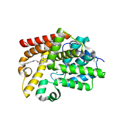 | | Crystal Structure of human phosphodiesterase 10 in complex with 2,3-dimethyl-6-[(1-methyl-4-phenylimidazol-2-yl)methoxy]imidazo[1,2-b]pyridazine | | 分子名称: | (4R)-2,3-dimethyl-6-[(1-methyl-4-phenyl-1H-imidazol-2-yl)methoxy]imidazo[1,2-b]pyridazine, MAGNESIUM ION, ZINC ION, ... | | 著者 | Joseph, C, Groebke-Zbinden, K, Benz, J, Schlatter, D, Rudolph, M.G. | | 登録日 | 2022-01-21 | | 公開日 | 2022-10-12 | | 最終更新日 | 2024-04-03 | | 実験手法 | X-RAY DIFFRACTION (1.94 Å) | | 主引用文献 | A high quality, industrial data set for binding affinity prediction: performance comparison in different early drug discovery scenarios.
J.Comput.Aided Mol.Des., 36, 2022
|
|
5SGN
 
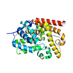 | | Crystal Structure of human phosphodiesterase 10 in complex with 2,3-dimethyl-6-[2-(2-methyl-5-pyrrolidin-1-yl-1,2,4-triazol-3-yl)ethyl]-7-(trifluoromethyl)imidazo[1,2-b]pyridazine | | 分子名称: | (4S)-2,3-dimethyl-6-{2-[1-methyl-3-(pyrrolidin-1-yl)-1H-1,2,4-triazol-5-yl]ethyl}-7-(trifluoromethyl)imidazo[1,2-b]pyridazine, MAGNESIUM ION, ZINC ION, ... | | 著者 | Joseph, C, Benz, J, Flohr, A, Groebke-Zbinden, K, Rudolph, M.G. | | 登録日 | 2022-02-01 | | 公開日 | 2022-10-12 | | 最終更新日 | 2024-04-03 | | 実験手法 | X-RAY DIFFRACTION (2.07 Å) | | 主引用文献 | Crystal Structure of a human phosphodiesterase 10 complex
To be published
|
|
5SH3
 
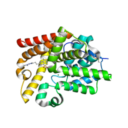 | | Crystal Structure of human phosphodiesterase 10 in complex with 2,3-dimethyl-6-[2-(2-methyl-5-pyrrolidin-1-yl-1,2,4-triazol-3-yl)ethyl]-8-methylsulfonylimidazo[1,2-b]pyridazine | | 分子名称: | (4S)-8-(methanesulfonyl)-2,3-dimethyl-6-{2-[1-methyl-3-(pyrrolidin-1-yl)-1H-1,2,4-triazol-5-yl]ethyl}imidazo[1,2-b]pyridazine, MAGNESIUM ION, ZINC ION, ... | | 著者 | Joseph, C, Benz, J, Flohr, A, Groebke-Zbinden, K, Rudolph, M.G. | | 登録日 | 2022-02-01 | | 公開日 | 2022-10-12 | | 最終更新日 | 2024-04-03 | | 実験手法 | X-RAY DIFFRACTION (1.98 Å) | | 主引用文献 | Crystal Structure of a human phosphodiesterase 10 complex
To be published
|
|
5SIB
 
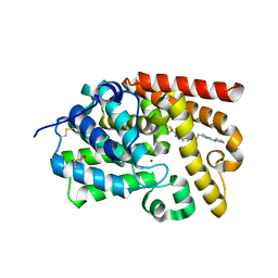 | | Crystal Structure of human phosphodiesterase 10 in complex with 3-methyl-6-[2-(2-methyl-5-pyrrolidin-1-yl-1,2,4-triazol-3-yl)ethyl]-2-(trifluoromethyl)imidazo[1,2-b]pyridazine | | 分子名称: | (4R)-3-methyl-6-{2-[1-methyl-3-(pyrrolidin-1-yl)-1H-1,2,4-triazol-5-yl]ethyl}-2-(trifluoromethyl)imidazo[1,2-b]pyridazine, MAGNESIUM ION, ZINC ION, ... | | 著者 | Joseph, C, Benz, J, Flohr, A, Groebke-Zbinden, K, Rudolph, M.G. | | 登録日 | 2022-02-01 | | 公開日 | 2022-10-12 | | 最終更新日 | 2024-04-03 | | 実験手法 | X-RAY DIFFRACTION (2.54 Å) | | 主引用文献 | Crystal Structure of a human phosphodiesterase 10 complex
To be published
|
|
3T19
 
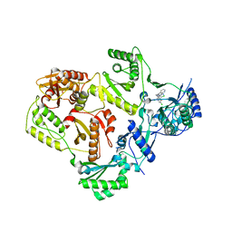 | |
3UP8
 
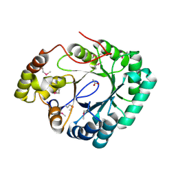 | | Crystal structure of a putative 2,5-diketo-D-gluconic acid reductase B | | 分子名称: | ACETATE ION, Putative 2,5-diketo-D-gluconic acid reductase B | | 著者 | Eswaramoorthy, S, Chamala, S, Evans, B, Foti, R, Gizzi, A, Hillerich, B, Kar, A, LaFleur, J, Seidel, R, Villigas, G, Zencheck, W, Almo, S.C, Swaminathan, S, New York Structural Genomics Research Consortium (NYSGRC) | | 登録日 | 2011-11-17 | | 公開日 | 2011-12-14 | | 最終更新日 | 2023-12-06 | | 実験手法 | X-RAY DIFFRACTION (1.96 Å) | | 主引用文献 | Crystal structure of a putative 2,5-diketo-D-gluconic acid reductase B
To be Published
|
|
