Loading
PDBj
メニューPDBj@FacebookPDBj@TwitterPDBj@YouTubewwPDB FoundationwwPDB
RCSB PDBPDBeBMRBAdv. SearchSearch help

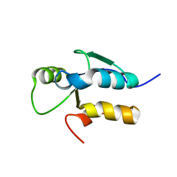
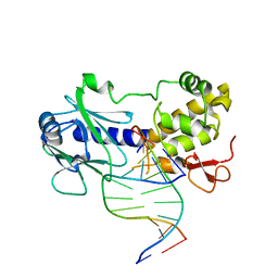
3JR4
DownloadVisualize
BU of 3jr4 by Molmil
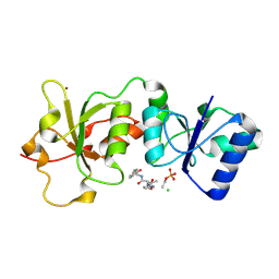
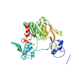
MutM interrogating an extrahelical G
分子名称: DNA (5'-D(*AP*GP*GP*TP*AP*GP*AP*CP*CP*TP*GP*GP*AP*CP*GP*C)-3'), DNA (5'-D(*TP*G*CP*GP*TP*CP*CP*AP*(GX1)P*GP*TP*CP*TP*AP*CP*C)-3'), DNA glycosylase, ...
著者Qi, Y, Spong, M.C, Verdine, G.L.
登録日2009-09-08
公開日2009-11-03
最終更新日2023-09-06
実験手法X-RAY DIFFRACTION (2.601 Å)
主引用文献Entrapment and structure of an extrahelical guanine attempting to enter the active site of a bacterial DNA glycosylase, MutM.
J.Biol.Chem., 285, 2010
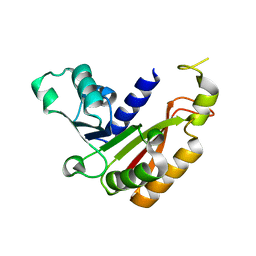
3JR9
DownloadVisualize
BU of 3jr9 by Molmil
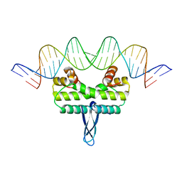
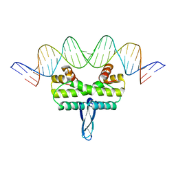
Crystal structure of Fis bound to 27 bp optimal binding sequence F2
分子名称: DNA (27-MER), DNA-binding protein fis
著者Stella, S, Cascio, D, Johnson, R.C.
登録日2009-09-08
公開日2010-04-28
最終更新日2023-09-06
実験手法X-RAY DIFFRACTION (2.9 Å)
主引用文献The shape of the DNA minor groove directs binding by the DNA-bending protein Fis.
Genes Dev., 24, 2010
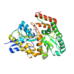
3JRF
DownloadVisualize
BU of 3jrf by Molmil
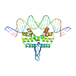
Crystal structure of Fis bound to 27 bp DNA F27 containing a C/G at center
分子名称: DNA (27-MER), DNA-binding protein fis
著者Stella, S, Cascio, D, Johnson, R.C.
登録日2009-09-08
公開日2010-04-28
最終更新日2024-02-21
実験手法X-RAY DIFFRACTION (3.05 Å)
主引用文献The shape of the DNA minor groove directs binding by the DNA-bending protein Fis.
Genes Dev., 24, 2010
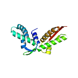
3JRP
DownloadVisualize
BU of 3jrp by Molmil
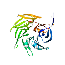
SEC13 with NUP145C (AA109-179) insertion blade
分子名称: FUSION PROTEIN OF PROTEIN TRANSPORT PROTEIN SEC13 AND NUCLEOPORIN NUP145
著者Brohawn, S.G, Gogola, M, Schwartz, T.U.
登録日2009-09-08
公開日2009-10-27
最終更新日2023-09-06
実験手法X-RAY DIFFRACTION (2.6 Å)
主引用文献Molecular architecture of the Nup84-Nup145C-Sec13 edge element in the nuclear pore complex lattice.
Nat.Struct.Mol.Biol., 16, 2009
3JSK
DownloadVisualize
BU of 3jsk by Molmil
Thiazole synthase from Neurospora crassa
分子名称: ADENOSINE DIPHOSPHATE 5-(BETA-ETHYL)-4-METHYL-THIAZOLE-2-CARBOXYLIC ACID, CyPBP37 protein, FE (II) ION
著者Kang, Y.N, Bale, S, Ealick, S.E.
登録日2009-09-10
公開日2010-09-29
最終更新日2023-09-06
実験手法X-RAY DIFFRACTION (2.7 Å)
主引用文献Crystal structure of thiazole synthase Thi4 from Neurospora crassa
to be published
3JUA
DownloadVisualize
BU of 3jua by Molmil
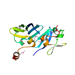
Structural basis of YAP recognition by TEAD4 in the Hippo pathway
分子名称: 65 kDa Yes-associated protein, Transcriptional enhancer factor TEF-3
著者Chen, L, Song, H.
登録日2009-09-15
公開日2010-02-23
最終更新日2024-03-20
実験手法X-RAY DIFFRACTION (3 Å)
主引用文献Structural basis of YAP recognition by TEAD4 in the hippo pathway.
Genes Dev., 24, 2010
3JUJ
DownloadVisualize
BU of 3juj by Molmil
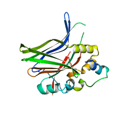
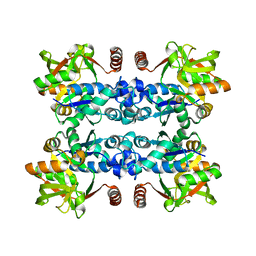
The crystal structure of apo- UDP-glucose pyrophosphorylase
分子名称: UDP-glucose pyrophosphorylase (GalU)
著者Kim, H, Kim, K.K.
登録日2009-09-15
公開日2010-03-31
最終更新日2024-03-20
実験手法X-RAY DIFFRACTION (2.9 Å)
主引用文献Structural basis for the reaction mechanism of UDP-glucose pyrophosphorylase
Mol.Cells, 29, 2010
3JV0
DownloadVisualize
BU of 3jv0 by Molmil
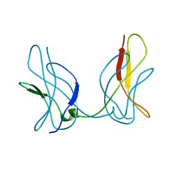
Crystal structure of a mutant of RelB dimerization domain(M6)
分子名称: Transcription factor RelB
著者Vu, D, Huang, D.B, Ghosh, G.
登録日2009-09-15
公開日2010-11-24
最終更新日2023-09-06
実験手法X-RAY DIFFRACTION (2.65 Å)
主引用文献Instability of the RelB dimerization domain is functionally importent
To be Published
3JV5
DownloadVisualize
BU of 3jv5 by Molmil
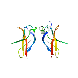
Crystal structure of the dimerization domains p52 homodimer
分子名称: Nuclear factor NF-kappa-B p100 subunit
著者Vu, D, Huang, D.B, Ghosh, G.
登録日2009-09-15
公開日2010-11-24
最終更新日2024-02-21
実験手法X-RAY DIFFRACTION (2.65 Å)
主引用文献Instability of the RelB dimerization domain is functionally important
To be Published
3JVE
DownloadVisualize
BU of 3jve by Molmil
Crystal Structure of the Sixth BRCT Domain of TopBP1
分子名称: DNA topoisomerase 2-binding protein 1
著者Leung, C.C, Kellogg, E, Baker, D, Glover, J.N.M.
登録日2009-09-16
公開日2010-01-19
最終更新日2024-02-21
実験手法X-RAY DIFFRACTION (1.34 Å)
主引用文献Insights from the crystal structure of the sixth BRCT domain of topoisomerase IIbeta binding protein 1.
Protein Sci., 19, 2010
3JWI
DownloadVisualize
BU of 3jwi by Molmil
Crystal structure analysis of the methyltransferase domain of bacterial-CtHen1-CN
分子名称: Methyltransferase type 12
著者Huang, R.H, Chan, C.M, Zhou, C, Brunzelle, J.S.
登録日2009-09-18
公開日2009-10-20
最終更新日2024-02-21
実験手法X-RAY DIFFRACTION (2.2 Å)
主引用文献Structural and biochemical insights into 2'-O-methylation at the 3'-terminal nucleotide of RNA by Hen1.
Proc.Natl.Acad.Sci.USA, 106, 2009
3K02
DownloadVisualize
BU of 3k02 by Molmil
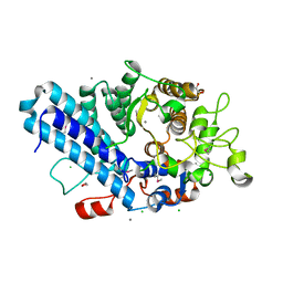
Crystal structures of the GacH receptor of Streptomyces glaucescens GLA.O in the unliganded form and in complex with acarbose and an acarbose homolog. Comparison with acarbose-loaded maltose binding protein of Salmonella typhimurium.
分子名称: 4,6-dideoxy-4-{[(1S,2R,3R,4S,5S)-2,3,4-trihydroxy-5-(hydroxymethyl)cyclohexyl]amino}-alpha-D-allopyranosyl-(1->4)-alpha-D-glucopyranosyl-(1->4)-alpha-D-glucopyranosyl-(1->4)-alpha-D-glucopyranose, Acarbose/maltose binding protein GacH, SULFATE ION
著者Vahedi-Faridi, A, Licht, A, Bulut, H, Schneider, E.
登録日2009-09-24
公開日2010-02-16
最終更新日2024-02-21
実験手法X-RAY DIFFRACTION (1.55 Å)
主引用文献Crystal Structures of the Solute Receptor GacH of Streptomyces glaucescens in Complex with Acarbose and an Acarbose Homolog: Comparison with the Acarbose-Loaded Maltose-Binding Protein of Salmonella typhimurium.
J.Mol.Biol., 397, 2010
3K10
DownloadVisualize
BU of 3k10 by Molmil
Crystal structure of telomere capping protein Stn1 from Saccharomyces cerevisiae
分子名称: Protein STN1
著者Gelinas, A.D, Reyes, F.E, Batey, R.T, Wuttke, D.S.
登録日2009-09-25
公開日2009-10-27
最終更新日2017-11-01
実験手法X-RAY DIFFRACTION (2.5 Å)
主引用文献Telomere capping proteins are structurally related to RPA with an additional telomere-specific domain.
Proc.Natl.Acad.Sci.USA, 106, 2009
3K16
DownloadVisualize
BU of 3k16 by Molmil
Crystal Structure of BRCA1 BRCT D1840T in complex with a minimal recognition tetrapeptide with a free carboxy C-terminus
分子名称: Breast cancer type 1 susceptibility protein, CHLORIDE ION, NICKEL (II) ION, ...
著者Campbell, S.J, Edwards, R.A, Glover, J.N.
登録日2009-09-25
公開日2010-03-02
最終更新日2021-10-13
実験手法X-RAY DIFFRACTION (3 Å)
主引用文献Comparison of the Structures and Peptide Binding Specificities of the BRCT Domains of MDC1 and BRCA1
Structure, 18, 2010
3JPV
DownloadVisualize
BU of 3jpv by Molmil
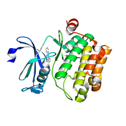
Crystal structure of human proto-oncogene serine threonine kinase (PIM1) in complex with a consensus peptide and a pyrrolo[2,3-a]carbazole ligand
分子名称: 1,10-dihydropyrrolo[2,3-a]carbazole-3-carbaldehyde, Peptide (PIMTIDE) ARKRRRHPSGPPTA, Proto-oncogene serine/threonine-protein kinase Pim-1
著者Filippakopoulos, P, Bullock, A.N, Fedorov, O, Akue-Gedu, R, Rossignol, E, Azzaro, S, Bain, J, Cohen, P, Prudhomme, M, Moreau, P, Amizon, F, von Delft, F, Arrowsmith, C.H, Weigelt, J, Edwards, A, Bountra, C, Knapp, S, Structural Genomics Consortium (SGC)
登録日2009-09-04
公開日2009-10-27
最終更新日2023-09-06
実験手法X-RAY DIFFRACTION (2.35 Å)
主引用文献Synthesis, kinase inhibitory potencies, and in vitro antiproliferative evaluation of new pim kinase inhibitors.
J.Med.Chem., 52, 2009
3JQ1
DownloadVisualize
BU of 3jq1 by Molmil
Crystal structure of SusD superfamily protein (YP_001297730.1) from Bacteroides vulgatus ATCC 8482 at 1.55 A resolution
分子名称: 1,2-ETHANEDIOL, CALCIUM ION, CHLORIDE ION, ...
著者Joint Center for Structural Genomics (JCSG)
登録日2009-09-04
公開日2009-09-22
最終更新日2023-02-01
実験手法X-RAY DIFFRACTION (1.55 Å)
主引用文献Crystal structure of SusD superfamily protein (YP_001297730.1) from Bacteroides vulgatus ATCC 8482 at 1.55 A resolution
To be published
3JQC
DownloadVisualize
BU of 3jqc by Molmil
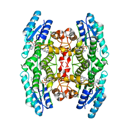
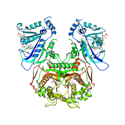
Crystal structure of pteridine reductase 1 (PTR1) from Trypanosoma brucei in ternary complex with cofactor (NADP+) and inhibitor 2-amino-6-bromo-4-oxo-4,7-dihydro-3H-pyrrolo[2,3-d]pyrimidine-5-carbonitrile (JU2)
分子名称: 2-amino-6-bromo-4-oxo-4,7-dihydro-3H-pyrrolo[2,3-d]pyrimidine-5-carbonitrile, NADP NICOTINAMIDE-ADENINE-DINUCLEOTIDE PHOSPHATE, Pteridine reductase 1
著者Tulloch, L.B, Hunter, W.N.
登録日2009-09-06
公開日2009-12-08
最終更新日2023-09-06
実験手法X-RAY DIFFRACTION (1.8 Å)
主引用文献Structure-based design of pteridine reductase inhibitors targeting african sleeping sickness and the leishmaniases.
J.Med.Chem., 53, 2010
3K50
DownloadVisualize
BU of 3k50 by Molmil
Crystal structure of Putative S41 protease (YP_211611.1) from Bacteroides fragilis NCTC 9343 at 2.00 A resolution
分子名称: CHLORIDE ION, GLYCEROL, Putative S41 protease
著者Joint Center for Structural Genomics (JCSG)
登録日2009-10-06
公開日2009-10-13
最終更新日2023-02-01
実験手法X-RAY DIFFRACTION (2 Å)
主引用文献Crystal structure of Putative S41 protease (YP_211611.1) from Bacteroides fragilis NCTC 9343 at 2.00 A resolution
To be published
3K7A
DownloadVisualize
BU of 3k7a by Molmil
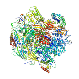
Crystal Structure of an RNA polymerase II-TFIIB complex
分子名称: DNA-directed RNA polymerase II subunit RPB1, DNA-directed RNA polymerase II subunit RPB11, DNA-directed RNA polymerase II subunit RPB2, ...
著者Liu, X, Bushnell, D.A, Wang, D, Calero, G, Kornberg, R.D.
登録日2009-10-12
公開日2009-11-24
最終更新日2023-09-06
実験手法X-RAY DIFFRACTION (3.8 Å)
主引用文献Structure of an RNA polymerase II-TFIIB complex and the transcription initiation mechanism.
Science, 327, 2010
3JRD
DownloadVisualize
BU of 3jrd by Molmil
Crystal structure of Fis bound to 27 bp DNA F25 containing T2A3 sequence at center
分子名称: DNA (27-MER), DNA-binding protein fis
著者Stella, S, Cascio, D, Johnson, R.C.
登録日2009-09-08
公開日2010-04-28
最終更新日2023-09-06
実験手法X-RAY DIFFRACTION (3.1 Å)
主引用文献The shape of the DNA minor groove directs binding by the DNA-bending protein Fis.
Genes Dev., 24, 2010
3JRM
DownloadVisualize
BU of 3jrm by Molmil
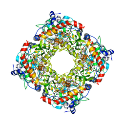
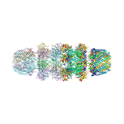
Crystal structure of archaeal 20S proteasome in complex with mutated P26 activator
分子名称: Proteasome activator protein PA26, Proteasome subunit alpha, Proteasome subunit beta
著者Stadtmueller, B.M, Whitby, F.G, Hill, C.P.
登録日2009-09-08
公開日2009-11-03
最終更新日2023-09-06
実験手法X-RAY DIFFRACTION (2.9 Å)
主引用文献Structural models for interactions between the 20S proteasome and its PAN/19S activators.
J.Biol.Chem., 285, 2010
3K8R
DownloadVisualize
BU of 3k8r by Molmil
Crystal structure of protein of unknown function (YP_427503.1) from Rhodospirillum rubrum ATCC 11170 at 2.75 A resolution
分子名称: 1,2-ETHANEDIOL, Uncharacterized protein
著者Joint Center for Structural Genomics (JCSG)
登録日2009-10-14
公開日2009-10-27
最終更新日2023-02-01
実験手法X-RAY DIFFRACTION (2.75 Å)
主引用文献Crystal structure of protein of unknown function (YP_427503.1) from Rhodospirillum rubrum ATCC 11170 at 2.75 A resolution
To be published
3JSU
DownloadVisualize
BU of 3jsu by Molmil
Quadruple mutant(N51I+C59R+S108N+I164L) plasmodium falciparum dihydrofolate reductase-thymidylate synthase(PFDHFR-TS) complexed with QN254, NADPH, and dUMP
分子名称: 2'-DEOXYURIDINE 5'-MONOPHOSPHATE, 5-chloro-N~6~-(2,5-dimethoxybenzyl)quinazoline-2,4,6-triamine, Dihydrofolate reductase-thymidylate synthase, ...
著者Chitnumsub, P, Maneeruttanarungroj, C, Kamchonwongpaisan, S, Yuthavong, Y, Diagana, T.T.
登録日2009-09-11
公開日2010-07-28
最終更新日2023-11-01
実験手法X-RAY DIFFRACTION (2.7 Å)
主引用文献Preclinical evaluation of the antifolate QN254, 5-chloro- N'6'-(2,5-dimethoxy-benzyl)-quinazoline-2,4,6-triamine, as an antimalarial drug candidate
Antimicrob.Agents Chemother., 54, 2010
3K8Y
DownloadVisualize
BU of 3k8y by Molmil
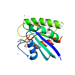
Allosteric modulation of H-Ras GTPase
分子名称: ACETATE ION, CALCIUM ION, GTPase HRas, ...
著者Holzapfel, G, Buhrman, G, Mattos, C.
登録日2009-10-15
公開日2010-03-02
最終更新日2023-09-06
実験手法X-RAY DIFFRACTION (1.3 Å)
主引用文献Allosteric modulation of Ras positions Q61 for a direct role in catalysis.
Proc.Natl.Acad.Sci.USA, 107, 2010
3JTX
DownloadVisualize
BU of 3jtx by Molmil
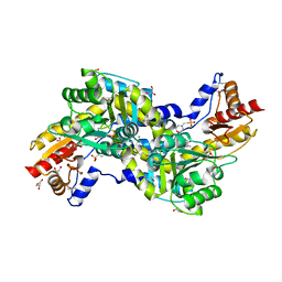
Crystal structure of Aminotransferase (NP_283882.1) from NEISSERIA MENINGITIDIS Z2491 at 1.91 A resolution
分子名称: 2-(N-MORPHOLINO)-ETHANESULFONIC ACID, ACETATE ION, CALCIUM ION, ...
著者Joint Center for Structural Genomics (JCSG)
登録日2009-09-14
公開日2009-09-22
最終更新日2023-02-01
実験手法X-RAY DIFFRACTION (1.91 Å)
主引用文献Crystal structure of Aminotransferase (NP_283882.1) from NEISSERIA MENINGITIDIS Z2491 at 1.91 A resolution
To be published

224201

件を2024-08-28に公開中

PDB statisticsPDBj update infoContact PDBjnumon